A crucial developmental factor in progression of mammalian
cerebral cortex which lead to microdeletion genetic disorder
INTRODUCTION
Chromosome 15q24 microdeletion syndrome is a well-described rare microdeletion genetic disorder that affects growth and development. Individuals with this condition are typically characterized by pre and post-natal growth retardation, intellectual disabilities, and unique facial features. They may also have abnormalities in their skeletal
structure, digits, and genitalia. Other common characteristics include low muscle tone (hypotonia), behavioral challenges, and frequent infections.
15q24 deletion syndrome occurs when a small section of chromosome 15, specifically the 15q24 region, is lost. This missing section can vary in size, typically ranging from about 1.7
to 6.1 million DNA base pairs (Mb). The deletion happens due to a process called nonallelic homologous recombination (NAHR), where the genetic material from similar regions of the chromosome, known as low-copy repeats (LCRs), misalign and lead to the loss of a portion
of the chromosome. They have also identified five key LCR clusters in the 15q24 region, labeled LCR15q24A,LCR15q24B, LCR15q24C, LCR15q24D, and LCR15q24E. These clusters act as hotspots for the deletion events, and depending on where the break occurs, it leads to different sizes of deletions, which in turn cause the symptoms of the syndrome.
INVOLVEMENT OF GENE
SIN3A play roles in tumor suppression and their deletion may lead to an increased predisposition to and risk of neoplasia in this patient population.
SIN3A gene encodes a protein similar to yeast Sin3A and is linked to Witteveen-Kolk syndrome (WITKOS), a condition inherited in an autosomal dominant manner. Individuals with WITKOS typically exhibit intellectual disabilities and distinct facial features.
Heterozygous truncating mutations in the SIN3A gene in patients with this syndrome. Studies on mice have shown that reducing Sin3a levels leads to fewer cortical progenitor neurons in the brain’s proliferative zone, indicating that SIN3A is crucial for the development of the mammalian cerebral cortex.
SYMPTOMS
Growth delay, feeding difficulties, and distinct facial features were often the presenting early symptoms of 15q24 deletion syndrome. Distinct facial features, including long face with high
anterior hairline, epicanthal folds, hypertelorism, downslanting palpebral fissures, sparse and broad medial eyebrows, broad and/or depressed nasal bridge, long smooth philtrum, and small
mouths with full lower lips were common in all individuals.
REPORTS
A recent study by Cooper et al. reviewed 15,767 cases with intellectual disability, however 16 of the 19 reported cases (84%) are males.
ANOTHER FINDING
Some patients have had breakpoints that do not lie within the LCR regions. Breakpoint sequencing for the 15q24 deletion in one of those patients revealed microhomology suggesting that other possible mechanisms include nonhomologous end joining (NHEJ) and
fork stalling and template switching (FoSTeS)/microhomology-mediated break-induced replication (MMBIR).
Acheiropodia is inherited in an autosomal recessive manner led to congenital disorder
INTRODUCTION
Limb–body wall complex also known as body stalk syndrome, is a rare congenital disorder marked by severe malformations affecting the limbs, thorax, and abdomen. It is characterized by conditions such as thoracoschisis, abdominoschisis, and various limb defects. Acheiropodia is inherited in an autosomal recessive manner, meaning that individuals who carry only one copy of the mutated gene (heterozygotes) do not show any symptoms and appear completely normal. Acheiropodia is a rare condition characterized by the congenital absence of both the upper and lower limbs, with the hands and feet missing due to underdevelopment (aplasia). This condition affects both sides of the body (bilateral).
INVOLVEMENT OF GENE
Mapping of the PPD locus on chromosome 7q36, which overlaps partially with the critical region for acheiropodia, identified three genes: C7orf2, C7orf3, and C7orf4. C7orf2 is the human equivalent of the mouse Lmbr1 gene and is thought to encode a receptor.
×
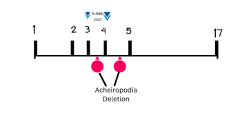
Fig : A composite representation of the human and mouse Lmbr1/LMBR1 genes illustrates the relative positions of limb mutations associated with these genes. The Lmbr1 gene, which is composed of 17 exons, is shown to be involved in various limb development mutations in both species. The acheiropodia deletion is located around exon 4, with the majority of the 5–6 kb deletion consisting of intronic DNA.
It is evident that C7orf2/Lmbr1 plays a vital role in the formation and development of distal limbs. The mouse Lmbr1 gene exhibits significant changes in expression in the limbs of Hemimelic extra toes (Hx) mice. The Hx mutation leads to hemimelia (underdevelopment) of the radius and tibia, along with preaxial polydactyly (extra digits) on both the forelimbs and hind limbs. Given the involvement of the Lmbr1 gene in limb abnormalities and its genomic location, C7orf2 is considered a strong candidate gene for acheiropodia in humans.
SYMPTOMS
Acheiropodia is a rare condition characterized by the congenital absence of both the upper and lower limbs, with the hands and feet missing due to underdevelopment (aplasia). This condition affects both sides of the body (bilateral).
REPORTS
This condition is extremely rare, with an incidence of 1 in every 14,000 to 31,000
pregnancies, according to large epidemiological studies. Many affected pregnancies end in miscarriage or stillbirth. Most infants who survive the prenatal period die shortly after birth.
ANOTHER FINDING
The predicted protein product of the Lmbr1/LMBR1 gene is a novel multipass transmembrane protein that does not fall into any known functional class but has been highly conserved in different organisms. Its structure suggests that it may encode a membrane anchoring protein, adhesion molecule, transporter, or cell surface receptor. An important goal for future studies will be to determine how this gene interacts with other pathways and how it acts to control the development of distal skeletal structures in the vertebrate limb.
Understanding Epilepsy: Insights, Challenges, and Support
What is Epilepsy?
Epilepsy is a neurological disorder characterized by the tendency to have recurrent seizures due to abnormal electrical activity in the brain. So, what is seizure, it is abnormal electrical activity in the brain which leads to abnormal experiences like abnormal sensory experiences, abnormal motor experiences and abnormal psychomotor experiences.
It can affect anyone, regardless of age, gender, or. Throughout history, epilepsy has been viewed with a mix of fear and misunderstanding, often attributed to supernatural causes. Thankfully, our understanding has evolved significantly, focusing more on its medical aspects and the various ways it can be managed.
It is important to clarify that epilepsy is not the same as having a seizure disorder. While all individuals with epilepsy experience seizures, not all seizure disorders qualify as epilepsy. Epilepsy is diagnosed when a person has had two or more unprovoked seizures.
Abnormal Sensory Experiences
When it comes to abnormal sensory experiences, the temporal lobes are often involved. These sensory disturbances, commonly associated with neurological conditions, including epilepsy, can manifest in several ways:
Paresthesia: A tingling or "pins and needles" sensation, often felt in the hands, feet, or other parts of the body. This sensation can be transient or chronic.
Visual Disturbances: Includes seeing flashing lights, colors, shapes, or even experiencing hallucinations. Some individuals may also experience temporary vision loss or blurriness
Auditory Disturbances: Hearing unusual sounds such as ringing (tinnitus), buzzing, voices, or music that isn’t there
Derealization/Depersonalization: A feeling of detachment from one's surroundings or body, like being in a dream or observing oneself from outside the body.
Tactile Hallucinations: Sensation of being touched, insects crawling on the skin, or other sensations without any physical cause.
Abnormal Motor Experiences
Abnormal motor experiences involve the frontal lobes and are characterized by involuntary movements or a loss of control over motor functions, often due to abnormal electrical activity in the brain. These experiences can vary widely depending on the type of seizure:
Paresthesia: A tingling or "pins and needles" sensation, often felt in the hands, feet, or other parts of the body. This sensation can be transient or chronic.
Visual Disturbances: Includes seeing flashing lights, colors, shapes, or even experiencing hallucinations. Some individuals may also experience temporary vision loss or blurriness
Auditory Disturbances: Hearing unusual sounds such as ringing (tinnitus), buzzing, voices, or music that isn’t there
Derealization/Depersonalization: A feeling of detachment from one's surroundings or body, like being in a dream or observing oneself from outside the body.
Tactile Hallucinations: Sensation of being touched, insects crawling on the skin, or other sensations without any physical cause.
Abnormal Psychomotor Experiences
Abnormal psychomotor experiences involve both the temporal and frontal lobes and can manifest as distinctive and involuntary sensory, emotional, or motor phenomena. These experiences reveal how seizures can disrupt normal brain function and result in unusual perceptions, unexpected behaviors, or intense emotional shifts:
Deja Vu: A sensation of familiarity with a situation that feels as though it has happened before, often occurring before a seizure.
Jamais Vu: The unsettling experience of something familiar suddenly feeling strange or unfamiliar.
Olfactory Hallucinations: Experiencing smells that aren’t present, typically linked to seizure activity in the brain.
Emotional Blunting: A reduced or muted emotional response, often occurring during or after a seizure.
Gustatory Hallucinations: The perception of nonexistent tastes, often associated with seizures.
Abnormal Sexual Experiences: Intrusive and unexpected sexual thoughts or sensations occurring during seizures.
Anger Bursts: Sudden, intense outbursts of anger that are difficult to control and may be related to seizures.
Mood Fluctuations: Rapid and unpredictable changes in mood, commonly associated with seizure activity.
Types of Seizures
Seizures are generally categorized into two main types: generalized seizures and focal seizures.
Generalized Seizures: These involve both sides of the brain from the onset. They include:
Tonic-Clonic Seizures: Known as grand mal seizures, characterized by muscle stiffening (tonic phase) followed by jerking movements (clonic phase).
Absence Seizures: Brief episodes of staring or "zoning out," often mistaken for daydreaming.
Focal Seizures: These start in one area of the brain and can be either simple (with awareness) or complex (without awareness). They can cause various symptoms, sometimes leading to alterations in sensation or movement.
Causes and Risk Factors
Epilepsy can arise from a variety of causes and risk factors:
Genetic Factors: Some types of epilepsy are linked to genetic mutations and can run in families.
External Influences:
Head injuries, particularly those incurred during accidents, and infections such as meningitis can increase the risk of developing epilepsy.
Environmental and Lifestyle Factors:
Stress, poor sleep, and excessive alcohol consumption may trigger seizures in some individuals.
Understanding epilepsy and its complexities is crucial for those affected and their families. With advancements in medical research and support, there is hope for better management and quality of life for those living with this challenging condition.
MONA PRIYA TIRKEY
RESEARCH ASSISTANT EXRNA THERAPEUTICS
Neonatal seizures: Long-term prognosis in infants
INTRODUCTION
Seizures in the neonatal period commonly result from an acute neurological injury, such as
stroke or hypoxic–ischaemic encephalopathy (HIE) but rarer causes can also be encountered
including inborn errors of metabolism or neonatal epilepsy syndromes. Classification of
seizures that includes the following as major seizure types: subtle, clonic, tonic, and
myoclonic. Subtle seizure phenomena include those alterations in neonatal behavior, motor
functions, and autonomic function that are easily overlooked and that are not characterized by
clonic, tonic, or myoclonic activity. Such seizures include certain ocular phenomena, oral-
buccal-lingual movements, peculiar limb movements, autonomic alterations, and apnea.
Subtle seizures appear to be more common in premature than in full-term infants, and some
subtle clinical phenomena in full-term infants are not associated with simultaneous EEG
seizure activity.
INVOLVEMENT OF GENE
Several genes have been identified as playing a role in neonatal seizures.
Notable genes include KCNQ2, KCNQ3, ARX, STXBP1, SLC25A22, CDKL5,
KCNT1, SCN2A, and SCN8A. These genes are associated with various types of
neonatal epilepsies, which can influence the severity and frequency of seizures in
affected infants.
TREATMENT
Neonatal seizures can be extremely refractory to conventional AEDs, especially those
associated with HIE. Early diagnosis should isolate metabolic or infectious causes and direct
care to correct the primary cause. Current practices include early treatment with
phenobarbital (doses ranging from 20–40 mg/kg),32 with phenytoin (20 mg/kg), or
fosphenytoin, and/or benzodiazepines such as lorazepam (0.05–0.1 mg/kg) as second-line
adjuvant therapy for refractory seizures. Although continuous video electroencephalogram
(EEG) is the gold standard for monitoring neonatal seizures, amplitude-integrated EEGs have
gained significant popularity in resource-limited settings. There is tremendous progress in the
automated seizure detection algorithm, including the availability of a fully convolutional
neural network using artificial machine learning (deep learning).
REPORTS
Irrespective of their classification as acute symptomatic events or as manifestations of a
neonatal-onset epilepsy, neonatal seizures occur with an estimated incidence of 1/1000 to
5/1000 live births in population-based studies but a 8.6/1000 rate in a NICU-based study, with
considerably higher rates for preterm. Recently, in an epidemiological study considering only
EEG-confirmed neonatal seizures, a 5.0/1000 incidence was reported for neonates of 31–36
weeks of gestation, 54.9/1.000 for those of 28–30 weeks of gestation, and 85.6/1000 for
neonates of < 28 weeks of gestation. Furthermore, the risk of neonatal seizures is increasing
with decreasing birth weight.
FUTURE SCOPE
Refractory neonatal seizures remain a significant clinical problem, and no new treatments for
this condition has been introduced for decades. Many new mechanisms and components of
neonatal seizures have been uncovered. These present important new possibilities for novel
therapeutic strategies in the population of neonates at risk for acute and long-term neurologic
damage from neonatal seizures. Clinical therapeutic trials in neonates would be greatly
improved if there were accurate biomarkers of acute and chronic therapeutic efficacy, yet
none exist other than the EEG.
Alternative medications that work similar
to valproic acid in Seizure & Epilepsy
There are several alternative medications that exhibit similar anticonvulsant properties to valproic acid, commonly used for managing seizures. These alternatives are often preferred, especially in particular populations such as pregnant women, due to the lower risk of teratogenic effects associated with them. Medications such as lamotrigine and levetiracetam have been shown to be less teratogenic than valproic acid, making them suitable substitutes.
Lamotrigine
Lamotrigine is recognized as a first-line alternative to valproic acid, particularly due to its lower risk of teratogenicity when used during pregnancy. Studies have indicated that lamotrigine is statistically less likely to cause birth defects compared to valproic acid. This makes lamotrigine a preferred choice for women of childbearing age who require effective seizure control.
Levetiracetam
Levetiracetam is another anticonvulsant that is frequently utilized as an alternative to valproic acid. It has demonstrated a favourable safety profile and is known to have a lower incidence of teratogenic effects. Levetiracetam is usually recommended when patients experience adverse effects from valproic acid or when there are concerns regarding pregnancy implications.
Importance of Medical Consulltation
It’s crucial for healthcare providers to evaluate the risk-benefit profile of these alternative medications in the context of a patient’s overall health and treatments. Each patient’s history,particularly for women of childbearing age, requires thorough counseling regarding the implications of antiepileptic drug use during pregnancy. This personalized approach helps in optimizing seizure management while minimizing potential risks to the patient and the fetus.
Consciousness and Epilepsy: What Causes the “Absence” in Absence Seizures?
INTRODUCTION
Absence seizures involve brief, sudden lapses of consciousness. They are more
common in children than in adults. A person having an absence seizure may
stare blankly into space for a few seconds. Then the person typically returns
quickly to being alert. Absence seizures, sometimes called petit mal seizures,
are brief, usually lasting less than 20 seconds. They cause symptoms that may
be barely noticeable. People having these seizures may appear like they’re
daydreaming or zoning out.
INVOLVEMENT OF GENE
The CACNA1H gene, encoding T-calcium channels, is linked to susceptibility
to absence epilepsy, particularly in populations like the Chinese Han and
Caucasians.
Sequencing was used to identify single-nucleotide polymorphisms (SNPs) and
mutations in the CACNA1H gene, which were analysed for their impact on
channel function and contribution to absence seizures.
REPORTS
About 50 per 100,000 children are diagnosed with epilepsy every year,
accounting for 25% of new epilepsy cases (2020). Up to 60% of children with
absence seizures.
FUTURE SCOPE
Expanding genetic studies to identify additional susceptibility genes beyond
CACNA1H could enhance our understanding of the genetic basis of absence
seizures. This could involve conducting genome-wide association studies
(GWAS) to discover new genetic variants linked to the condition.
Additionally, research could focus on developing targeted therapies that
specifically address neurotransmitter systems, such as the GABAergic and T-
calcium channels. Designing drugs that selectively modulate these pathways
may lead to more effective treatments with fewer side effects.
Dravet syndrome, also known as severe myoclonic epilepsy of infancy, is a rare and severe form of epilepsy that typically appears in an infant’s first year. It is characterized by frequent, prolonged seizures, developmental delays, growth issues, and mobility challenges. The seizures often start with a fever or warm temperatures and can last more than 5 minutes, with subsequent seizures of varying types.
The condition is caused by mutations in the SCN1A gene, which affects sodium ion movement in cells, though other genes can also be involved. Dravet syndrome affects about 1 in 15,700 people in the U.S. and is inherited in some cases but often arises spontaneously.
Diagnosis involves clinical evaluation, EEG, MRI, and genetic testing. There is no cure for Dravet syndrome, but treatments aim to reduce seizure frequency and severity. Options include medications such as clobazam and valproic acid, rescue medications, and potentially vagus nerve stimulation. Lifestyle changes, such as avoiding triggers like high temperatures, and a ketogenic diet may also help manage symptoms.
The prognosis includes lifelong management, with the possibility of severe complications and a higher risk of sudden unexpected death in epilepsy (SUDEP). Early diagnosis and intervention can improve outcomes, but ongoing support and monitoring are crucial.
Causes and Genetics
Genetic Mutations: The majority of Dravet syndrome cases (80-90%) involve mutations in the SCN1A gene, which encodes a sodium channel protein crucial for normal neuronal function. This mutation impairs the movement of sodium ions across neuronal membranes, disrupting normal electrical signaling in the brain and leading to seizures.
Spontaneous Mutations: While some individuals inherit the mutation from their parents, in many cases, the mutation occurs spontaneously during development. This means that even without a family history of the condition, a child can still develop Dravet syndrome
Symptoms and Progression
Early Signs: The condition typically presents with a prolonged tonic-clonic seizure, often triggered by a fever or hot environments, in an otherwise healthy infant.
Seizure Types: In addition to the initial tonic-clonic seizures, children with Dravet syndrome may experience various types of seizures, including focal seizures, myoclonic seizures, and atonic seizures.
Developmental Impact: Over time, seizures can lead to significant developmental delays and intellectual disability. Affected children may have issues with motor skills, speech, and coordination, and may display reduced muscle tone and an unsteady gait.
Autonomic Dysfunction: Dravet syndrome can affect the autonomic nervous system, leading to problems with body temperature regulation and other autonomic functions.
Diagnosis
Clinical Evaluation: Diagnosis is based on clinical symptoms and history of prolonged seizures.
EEG and Brain Imaging: Electroencephalography (EEG) and brain imaging (MRI) are used to assess seizure activity and rule out other conditions, although EEG results might not always show abnormalities early on.
Genetic Testing: Identifying mutations in the SCN1A gene through genetic testing can confirm the diagnosis, especially in infants with recurrent seizures.
Treatment and Management
Medications: Standard antiepileptic drugs may not be effective. Common treatments include clobazam and valproic acid, with additional options like topiramate, clonazepam, and others. Newer medications such as cannabidiol (Epidiolex), stiripentol (Diacomit), and fenfluramine (Fintepla) have been approved specifically for Dravet syndrome.
Rescue Medications: These are used for acute seizure management and include sedatives administered orally or via injection.
Vagus Nerve Stimulation (VNS): This therapy involves implanting a device that sends electrical pulses to the vagus nerve to help reduce seizure frequency.
Ketogenic Diet: This high-fat, low-carbohydrate diet can be effective in reducing seizure frequency in some individuals with Dravet syndrome.
Lifestyle Adjustments: Managing environmental factors such as avoiding overheating, high temperatures, and strenuous activities can help prevent seizures.
Prognosis and Support
Lifelong Condition: Dravet syndrome is a chronic condition with ongoing management needs. While medications and therapies can help control symptoms, seizures often persist.
Complications: The syndrome is associated with an increased risk of SUDEP, which often occurs during sleep. Careful monitoring is necessary, especially during bathing or swimming, due to an increased risk of drowning during seizures.
Support and Resources: Families may benefit from support groups and organizations such as the Dravet Syndrome Foundation, Epilepsy Foundation, and others that provide resources, advocacy, and connection with other families facing similar challenges.
Research and Future Directions
Ongoing Research: Scientists are actively researching potential treatments and cures by focusing on the genetic causes of Dravet syndrome, including the SCN1A gene. Advances in gene therapy and other novel treatments hold promise for the future.
This additional detail provides a comprehensive understanding of Dravet syndrome, covering its genetic basis, symptoms, diagnostic processes, treatment options, and support resources.
Juvenile myoclonic epilepsy (JME): When Will it End?
INTRODUCTION
Juvenile myoclonic epilepsy (JME) is a special syndrome within the primary
generalized epilepsies which is characterized clinically by irregular jerks of shoulders and arms (so-called impulsive petit mal) after awakening. Juvenile myoclonic epilepsy (JME) mutation generates linkage disequilibrium. Juvenile myoclonic epilepsy (JME) has long been considered to be the most common chronic idiopathic generalized epilepsy syndrome, requiring life-long antiepileptic drug (AED) therapy.
Seizures can be triggered by lack of sleep, stress, and flickering lights, like those from screens. Therefore, classic symptoms of epileptic seizures include: Muscle
jerks and contractions, Loss of consciousness, Weakness, usually in a specific
body part, A feeling of anxiety right before a seizure, Staring into space.
INVOLVEMENT OF GENE
Genes associated with JME include EFHC1, BRD2, CASR, mutations discovered in
the coding sequence of EFHC1 are heterozygous missense mutations.
TREATMENT:
These medicines include: Valproic acid, Levetiracetam, Lamotrigine, Clonazepam,
Topiramate. Valproic acid is the most effective drug for JME. But women shouldn’t
take it during their childbearing years. Valproic acid acts through multiple
mechanisms, including increased synthesis of gamma-aminobutyric acid (GABA) in the nerve terminals and decreased degradation of GABA at the level of the synapse.Valproic acid also has some ability to decrease excitation by glutamate and alter ionic conductance.
REPORTS
Estimated to be 1 per 100,000 persons, with a prevalence of 0.1 to 0.2 per 100,000.
FUTURE SCOPE
The challenge of maintaining remission after AED withdrawal highlights the lack of disease-modifying effects in current treatments. To overcome this, there is a critical need to develop anti-epileptogenic therapies. Understanding the high relapse rates in JME after AED withdrawal requires deeper insights into the abnormal development of neuronal connections and brain networks during brain maturation in ictogenesis and epileptogenesis.
Imagine a world where every day is unpredictable—where moments of joy are often interrupted by the sudden onset of a seizure, and where the future feels uncertain. This is the reality for those living with Lennox-Gastaut Syndrome (LGS), a rare and severe form of epilepsy that demands resilience, hope, and a community of support.
What is Lennox-Gastaut Syndrome?
Lennox-Gastaut Syndrome isn’t just another type of epilepsy—it’s a complex, multifaceted condition that typically begins in early childhood, usually between the ages of 2 and 8. Picture a young child playing one moment and then suddenly being overtaken by a seizure. This is the challenge of LGS: it brings a mix of seizure types that are often resistant to treatment, combined with cognitive and behavioral difficulties that can overshadow a child’s developmental progress.
LGS is like a storm with unpredictable weather patterns having prevalence rate of 26/100000 population —children can experience tonic seizures (muscle stiffening), atonic seizures (sudden loss of muscle tone, causing falls), and atypical absence seizures (brief lapses in awareness).
These seizures can strike multiple times a day, making life unpredictable. Alongside the seizures,many children with LGS face intellectual disabilities and developmental delays, which can make simple tasks daunting.
A slow spike-and-wave pattern on an EEG is a signature of LGS, helping to distinguish it from other epilepsy types.
The Many Faces of LGS: Causes and Origins
LGS doesn’t have one single cause—think of it as a puzzle with many pieces, some of which are still missing. The syndrome can be triggered by several factors, each contributing a different piece to the puzzle:
Brain Malformations: Sometimes, the brain’s structure itself is altered, leading to LGS.
Genetics: While genetic mutations can be a cause, many cases remain unexplained, adding to the syndrome’s mystery.
Infections or Trauma: Imagine the brain as a delicate ecosystem; infections or injuries can disrupt this balance, leading to LGS.
Evolution from Other Epilepsy Syndromes: LGS can also be the next chapter in the story of a child who previously had another form of epilepsy, such as infantile spasms.
Diagnosing LGS: Piecing Together the Puzzle
Diagnosing LGS is like being a detective—each clue, whether it’s a child’s seizure history or the results of an EEG, brings us closer to understanding the full picture.
Medical History: A detailed review of the child’s experiences, from their earliest
seizures to their developmental milestones.
EEG Testing: This is where we see the telltale signs—the slow spike-and-wave pattern that’s characteristic of LGS.
Imaging and Genetic Testing: These tests help uncover underlying causes, whether it’s a brain malformation or a genetic mutation
Treatment: Managing the Unmanageable
Managing LGS is like trying to calm a storm—it takes a combination of tools, creativity, and perseverance.
Medications: Finding the right mix of antiepileptic drugs (AEDs) can be a long and
difficult process, often requiring adjustments over time.
Dietary Therapies: The ketogenic diet, high in fats and low in carbohydrates, has been a beacon of hope for some, helping to reduce seizure frequency.
Vagus Nerve Stimulation (VNS): Imagine a device that sends regular pulses to the
brain, helping to keep the storm at bay.
Surgery: In severe cases, surgery can be an option—like rerouting a river to prevent flooding, certain surgical procedures can help reduce seizure severity.
Cannabidiol (CBD): The FDA-approved drug Epidiolex, derived from CBD, offers a
new frontier in the battle against LGS, bringing hope to many families.
Living with LGS: The Everyday Reality
Life with LGS is a journey marked by resilience. It’s about finding moments of joy amidst the challenges, leaning on a community of support, and never giving up hope. For families, every day is a balancing act—managing medications, attending therapy sessions, and advocating for their child’s needs in school and beyond.
Support Systems: The journey is made easier by connecting with others who understand the challenges of LGS, from healthcare professionals to support groups.
Adapting to Change: As children with LGS grow, their needs evolve, requiring constant adjustments to care strategies.
Advocacy: Educating others about LGS and pushing for more research is crucial for changing the future for those affected by this syndrome.
Looking Ahead: A Future of Hope
While LGS remains a complex and challenging condition, the future is not without hope.
Advances in research, new treatments, and a deeper understanding of the syndrome are lighting the way forward. The quest for more effective therapies and, ultimately, a cure continues, driven by the unwavering determination of families, researchers, and advocates.
Autosomal dominant nocturnal frontal lobe epilepsy ( ADNFLE) with a mutation in the CHRNA4 gene
INTRODUCTION
Autosomal dominant nocturnal frontal lobe epilepsy (ADNFLE) is an uncommon form of epilepsy that runs in families. This disorder causes seizures that usually occur at night (nocturnally) while an affected person is sleeping. Some people with ADNFLE also have seizures during the day.
INVOLVEMENT OF GENE
Mutations in the CHRNA4 genes can cause ADNFLE. These genes provide instructions for making different parts (subunits) of a larger molecule called a neuronal nicotinic acetylcholine receptor (nAChR). This receptor plays an important role in chemical signaling between nerve cells (neurons) in the brain.
SYMPTOMS:
The most common symptoms associated with an aura in people with ADNFLE are tingling, shivering, a sense of fear, dizziness (vertigo), and a feeling of falling or being pushed. Some affected people have also reported a feeling of breathlessness, overly fast breathing (hyperventilation), or choking
MANAGEMENT AND TREATMENT:
The treatment of choice for ADNFLE includes use of carbamazepine (200-1,000 mg/day). Carbamazepine abolishes seizures in 20% of cases, and gives significant relief (at least 50 % seizure reduction) in another 48%. Oxcarbamazepine, topiramate and acetazolamide (as add-on therapies) may also be used. Nicotine transdermal patches may be efficient in patients who are refractory to standard antiepileptic drugs.
PREVALENCE:
ADNFLE appears to be an uncommon form of epilepsy; its prevalence is unknown. This condition has been reported in more than 100 families worldwide.
FUTURE SCOPE
The detailed contribution of these finding to ADNFLE pathogenesis is still poorly understood. In addition to the further research to find the consequences of developmental defects and cause of excessive glutamate transmission, more studies should be focused on the role of nicotine exposure during development and low doses of nicotine as a potent therapy. Nicotine has already been shown to be effective in a few patients, and more data on the therapeutic effects in relation to specific mutations are needed to establish nicotine as a new treatment and successor of carbamazepine
Early onset seizure in Atypical Rett syndrome: CDKL5 deficiency disorder
INTRODUCTION
Atypical RTT is a genetic neurodevelopmental disorder. It causes a child to develop both physical and mental disabilities. Atypical Rett Syndrome is diagnosed when a patient has at least two main features of classic Rett syndrome and a period of regression followed by stabilization or recovery. They must also meet five of eleven supportive criteria, such as breathing issues and abnormal muscle tone.
INVOLVEMENT OF GENE
The early-onset seizure type (Hanefeld variant) is frequently caused by mutations in the X-linked CDKL5 gene (Xp22). CDKL5 mutations are ongoing, with studies indicating that alternative splicing of the pre-mRNA plays a significant role in producing functional isoforms of the CDKL5 protein, further influencing its efficacy in neuronal signaling. CDKL5(107), which consists of a 960-amino acid protein that includes the N-terminal kinase domain but has a different C-terminus compared to the full-length isoform CDKL5(115)
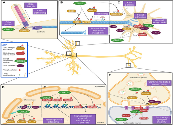
FIG: Known CDKL5 targets and affected processes in neurons. (https://doi.org/10.1042%2FBST20220791) CDKL5(107) shows highest expression levels in the brain, while CDKL5(115) is predominantly expressed in the testis. Both isoforms have been detected across various human tissues and cell lines, underscoring their potential importance in different physiological contexts
REPORT:
Based on reports that up to 32% of RTT cases show an atypical phenotype, the prevalence of atypical RTT is estimated at around 1/45,000. Like classic RTT, atypical RTT syndrome predominantly affects girls.
MAJOR FINDING:
A thorough understanding of the molecular pathways regulated by CDKL5 could uncover druggable targets, potentially accelerating the development of targeted therapies that directly address the underlying causes of CDKL5 Deficiency Disorder (CDD). Insights into CDKL5 signaling pathways not only promise to benefit patients with pathogenic CDKL5 variants but also have broader implications for understanding neurodegenerative disorders. Specifically, they highlight the crucial role of kinase-regulated signaling pathways in the brain. By translating these fundamental research findings into clinical applications, we have the potential to swiftly develop new therapeutics, profoundly improving the health of children who otherwise face a devastating and progressive disease trajectory due to CDKL5 deficiency.
Anti-epileptic drugs (AEDs), also known as anticonvulsants, are medications used to manage and prevent seizures in individuals with epilepsy. These drugs work by stabilizing electrical activity in the brain, thereby reducing the frequency and severity of seizures. The choice of AED depends on the type of epilepsy, the patient's age, health status, and other factors.
All anti- epileptic drugs are work on some mechanism of actions, the mechanism of actions are:
Prolongation of Na+ (Sodium ion) channel inactivation.
Facilitation of GABA (Gamma- Anti Butyric Acid) mediated Cl- (chlorine ion) channel opening.
Decrease in excitatory neurotransmission.
Inhibition of T- type Ca2+ channels.
The mechanism of action of anti-epileptic drugs (AEDs) involves various pathways and targets in the brain, all aimed at reducing neuronal excitability and preventing the abnormal electrical activity that leads to seizures. Here’s a detailed look at how different classes of AEDs work:
Prolongation of Na+ (Sodium ion) channel inactivation
Anti-epileptic drugs that prolong sodium channel inactivation work by Binding to sodium channels and keeping them in their inactivated state longer than usual than Reducing the ability of neurons to fire rapidly and repetitively which lowers neuronal excitability and preventing the occurrence of seizures.
This mechanism is particularly effective in managing various types of epilepsy, where controlling hyperactive neuronal circuits is crucial for preventing seizures.
Facilitation of GABA (Gamma- Anti Butyric Acid) mediated Cl- (chlorine ion) channel opening.
Antiepileptic drugs (AEDs) that enhance GABAergic activity work by facilitating the action of GABA, the brain's primary inhibitory neurotransmitter. These drugs increase the opening of chloride (Cl⁻) channels linked to GABA_A receptors. When GABA binds to these receptors, the Cl⁻ channels open, allowing Cl⁻ ions to flow into the neuron. This influx of Cl⁻ ions hyperpolarizes the neuron, making it less likely to fire. AEDs may enhance this process by increasing GABA release, boosting GABA receptor sensitivity, or prolonging the duration that Cl⁻ channels remain open. This increased inhibition helps reduce excessive neuronal firing and control seizures.
Decrease in excitatory neurotransmission:
Antiepileptic drugs (AEDs) that decrease excitatory neurotransmission work by reducing the brain’s excitatory signals. They achieve this by inhibiting the release of glutamate, the main excitatory neurotransmitter, which reduces neuronal stimulation. Some AEDs block excitatory receptors like AMPA or NMDA, preventing them from responding to glutamate. Additionally, certain drugs stabilize ion channels involved in generating excitatory signals, such as sodium or calcium channels. By targeting these mechanisms, these AEDs help to lower neuronal activity and reduce the likelihood of seizures.
Inhibition of T- type Ca2+ channels:
Antiepileptic drugs that inhibit T-type Ca²⁺ channels, such as ethosuximide and, to a lesser extent, valproic acid, work by blocking these specific low-voltage activated channels. This inhibition reduces the influx of calcium ions into thalamic neurons, thereby decreasing their rhythmic burst firing. By modulating the excitability of thalamic neurons, these drugs also disrupt abnormal thalamocortical communication, which is crucial for controlling absence seizures and stabilizing abnormal brain activity.
Lists of drugs that works based on above mechanism:
Prolongation of Na+ (Sodium ion) channel inactivation
Facilitation of GABA mediated Chlorine ion channel opening
Amyotrophic Lateral Sclerosis (ALS): Pioneering Antisense Oligonucleotide Therapy for a Rare Neurodegenerative Disease
Amyotrophic Lateral Sclerosis (ALS), also known as Lou Gehrig's disease, is a devastating neurodegenerative disorder that progressively robs individuals of their ability to move, speak, and ultimately breathe. Affecting an estimated 2 in 100,000 people globally each year, ALS is classified as a rare disease, though its impact on patients and families is profound and widespread. Despite extensive research, effective treatment options have been limited, driving an urgent need for innovative therapies.
At the forefront of this battle is the emerging field of antisense oligonucleotide (ASO) therapy, offering new hope in the quest to treat this debilitating condition. Our organization is deeply committed to advancing ASObased treatments, harnessing the power of genetic medicine to target the root causes of ALS.
Understanding ALS: A Complex Neurodegenerative Disorder
ALS is characterized by the progressive degeneration of motor neurons in the brain and spinal cord. These neurons are responsible for transmitting signals from the brain to the muscles, enabling voluntary movements. As these neurons deteriorate, patients experience muscle weakness, spasticity, and eventually paralysis. While cognitive function is typically preserved, the loss of motor function is relentless, leading to death within 3 to 5 years of diagnosis for most patients.
The causes of ALS are multifactorial, with a combination of genetic and environmental factors at play. About 10% of cases are familial, linked to mutations in specific genes such as SOD1, C9orf72, and FUS. The remaining 90% are sporadic, with no clear hereditary component. Despite this variability, the underlying pathology involves the accumulation of toxic proteins, oxidative stress, mitochondrial dysfunction, and inflammation, all contributing to motor neuron death.
The Promise of Antisense Oligonucleotide Therapy in ALS
Antisense oligonucleotides (ASOs) represent a promising therapeutic approach for ALS by directly targeting the genetic mutations and molecular pathways implicated in the disease. ASOs are short, synthetic strands of nucleotides designed to bind to specific RNA sequences, modulating gene expression or correcting abnormal RNA splicing. This precision allows ASOs to silence harmful genes, promote the degradation of toxic proteins, or restore normal protein function
In the context of ALS, ASOs have shown particular promise in targeting known genetic mutations. For instance, ASOs targeting the SOD1 gene, which accounts for about 2% of all ALS cases, have demonstrated the ability to reduce the production of toxic SOD1 protein aggregates in preclinical models. Clinical trials with SOD1targeted ASOs, such as tofersen, have shown encouraging results, slowing disease progression in patients with SOD1 mutations.
Another promising target is the C9orf72 gene, which harbors an abnormal hexanucleotide repeat expansion responsible for up to 40% of familial ALS cases. ASOs designed to reduce the toxic RNA foci and dipeptide repeat proteins produced by this mutation are currently in development, with earlyphase trials indicating potential benefits.
Overcoming Challenges in ASOBased Therapy
While ASO therapy holds significant promise, several challenges remain. Delivering ASOs to the central nervous system (CNS) is complex, requiring intrathecal administration to bypass the bloodbrain barrier. Additionally, the longterm safety and efficacy of ASOs need further investigation, particularly in light of potential immune responses and offtarget effects.
To address these challenges, our organization is dedicated to refining ASO design, optimizing delivery methods, and conducting rigorous clinical trials. By focusing on precision medicine, we aim to develop ASOs that not only target the genetic underpinnings of ALS but also offer personalized treatment options tailored to individual patient profiles.
The Future of ALS Treatment: A New Era of Hope
The development of ASObased therapies marks a new era in the treatment of ALS, offering the possibility of slowing or even halting the progression of this relentless disease. As research advances, we are hopeful that ASOs will become a cornerstone of ALS treatment, transforming the lives of patients and their families.
Our organization remains at the forefront of this exciting field, committed to driving innovation and collaboration in the pursuit of effective treatments for ALS and other rare neurodegenerative diseases. By continuing to push the boundaries of genetic medicine, we aim to bring new hope to those affected by ALS, paving the way for a future where this devastating disease is no longer a death sentence but a manageable condition.
BowenConradi Syndrome: Understanding a Rare Genetic Disorder and the Urgent Need for Therapeutic Intervention
BowenConradi Syndrome (BCS) is a rare, autosomal recessive genetic disorder that affects multiple systems in the body, primarily leading to severe developmental delays and physical malformations. First described in the Hutterite population of North America, BowenConradi Syndrome has a particularly high prevalence in this group due to the founder effect—when a small population carries a rare mutation, leading to increased frequency of the disorder within that population.
Though rare globally, BowenConradi Syndrome presents significant challenges for affected families and highlights the urgent need for new and innovative therapeutic approaches, particularly those targeting the disease's underlying genetic cause. This article will explore the genetic basis of BCS, its clinical manifestations, and why there is a growing demand for focused treatment strategies.
What is BowenConradi Syndrome?
BowenConradi Syndrome is caused by mutations in the EMG1 gene, which plays an essential role in ribosome biogenesis, a critical cellular process involved in protein synthesis. This mutation leads to the improper formation of ribosomes, which in turn affects cell growth and function across multiple tissues, resulting in the wide array of developmental issues seen in BCS.
The clinical features of BCS are apparent from birth and are often severe. Common characteristics of the syndrome include:
Intrauterine growth restriction (IUGR): Babies with BCS are usually born with very low birth weights and often experience poor growth during pregnancy.
Microcephaly: Abnormally small head size due to improper brain development
Severe developmental delays: Affected infants display delayed or absent milestones in motor skills, cognitive function, and speech development.
Skeletal abnormalities: Including foot deformities, such as rocker bottom feet, and other physical malformations like clinodactyly (curved fingers).
Facial dysmorphisms: Such as a prominent nose, small jaw (micrognathia), and abnormal eye features.
Feeding difficulties: Infants may have difficulty swallowing, which contributes to poor growth and malnutrition.
Unfortunately, many infants with BowenConradi Syndrome do not survive beyond infancy due to severe medical complications such as infections, failure to thrive, and respiratory issues.
The Genetic Cause: EMG1 Mutation
The root cause of BowenConradi Syndrome lies in the mutation of the EMG1 gene, located on chromosome 12. This gene encodes a protein that is crucial for the formation and function of ribosomes, the cellular machinery responsible for producing proteins. Ribosomes are essential for all cells, particularly during rapid growth and development, making them critical for fetal and infant development.
The EMG1 mutation in BCS disrupts ribosome biogenesis, causing widespread cellular dysfunction. As a result, growth and development are severely impaired, leading to the characteristic features of BowenConradi Syndrome. Because the condition is autosomal recessive, both parents must carry one copy of the mutated gene for a child to be affected, with a 25% chance of inheritance if both parents are carriers.
Why BowenConradi Syndrome is Severe and LifeThreatening
One of the reasons BowenConradi Syndrome is particularly devastating is due to the vital role of the EMG1 gene in all cells of the body. Ribosomes are involved in producing proteins that are necessary for nearly every biological process, so a defect in ribosome production affects multiple organ systems. This is why individuals with BCS experience such widespread, systemic issues, including poor brain development, skeletal malformations, and critical feeding and respiratory problems.
In most cases, infants with BowenConradi Syndrome pass away in the first few months of life, often from complications related to respiratory failure, infection, or failure to thrive due to feeding difficulties. The severity of the disease and the lack of curative treatments make BCS one of the most urgent rare diseases requiring focused research and therapeutic development.
The Need for Therapeutic Intervention: Exploring GeneTargeted Therapies
Given the severity of BowenConradi Syndrome and the lack of effective treatments, there is a pressing need for the development of novel therapies that can target the genetic root of the disease. Traditional treatments are primarily focused on managing symptoms—such as addressing feeding difficulties or providing respiratory support—but these measures do not address the underlying cause of the condition and offer limited improvement in life expectancy or quality of life.
Recent advances in gene based therapies, particularly antisense oligonucleotide (ASO) technology and gene editing approaches, provide a promising avenue for treating the genetic defects responsible for diseases like BowenConradi Syndrome. These therapies could offer the ability to directly target and correct the EMG1 mutation, potentially restoring normal ribosome function and improving cellular growth and development.
Here’s why gene based therapies could be a gamechanger for BowenConradi Syndrome:
Targeting the Cause, Not the Symptoms: By targeting the underlying genetic mutation in the EMG1 gene, therapies like ASOs could offer a more precise and effective treatment than conventional approaches that only manage symptoms.
Potential for Early Intervention: Since BowenConradi Syndrome often manifests in utero, early identification through genetic screening could allow for early therapeutic intervention, potentially before the most severe manifestations of the disease take hold.
Customizability for Rare Diseases: Gene targeted therapies can be customized to address the specific mutation present in an individual, offering a precision medicine approach that is particularly important for rare diseases like BowenConradi Syndrome.
Challenges in Developing Therapies for BowenConradi Syndrome
Despite the promise of gene based treatments, there are still challenges to overcome before these therapies can become a reality for Bowen Conradi Syndrome:
Limited Research and Resources:As with many rare diseases, BowenConradi Syndrome has not received extensive attention from the scientific community, leading to a lack of research funding and clinical trials. Without more investment in research, the development of therapies for BCS remains a distant goal.
Delivery Mechanisms: Delivering gene therapies to the correct tissues—especially in a condition like BCS that affects many organ systems—remains a major hurdle. Ensuring that a treatment reaches the cells where it's needed most is a complex challenge.
Ethical Considerations of Prenatal Treatment:Because BCS manifests so early, treating it before birth may offer the best outcomes. However, this raises ethical and practical concerns about fetal therapy, including the risks and the potential impact on both the mother and the developing baby.
A Call for Action: Supporting Rare Disease Research
BowenConradi Syndrome, like many rare genetic disorders, highlights the urgent need for increased research and development of therapies that target the genetic causes of these diseases. By focusing on the underlying mutation in the EMG1 gene, we can work towards a future where infants born with BowenConradi Syndrome have a chance not only to survive but to thrive.
At our organization, our mission is to drive forward innovative research into rare genetic diseases like BowenConradi Syndrome. By developing and supporting gene targeted therapies, we aim to address the root causes of these conditions and bring hope to families affected by severe, life threatening diseases. Every step forward in research brings us closer to lifesaving treatments for rare diseases that have long been overlooked.
In the meantime, our efforts to raise awareness about the severity and impact of BowenConradi Syndrome are vital. We must continue to advocate for increased research funding, support for affected families, and the development of new therapies that could change the course of this devastating disease.
Ankita Sharma
RESEARCH ASSISTANT EXRNA THERAPEUTICS
Unraveling Ataxia-Hypogonadism-Choroidal Dystrophy Syndrome: Insights Into a Rare Neurodegenerative Disorder
Introduction
Ataxia-hypogonadism-choroidal dystrophy syndrome—or Boucher-Neuhauser syndrome (BNHS)—is a sporadic neurodegenerative disorder with a slow, progressive course. This autosomal recessive condition is notable for its triad of hallmark features: cerebellar ataxia, chorioretinal dystrophy, and hypogonadotropic hypogonadism. BNHS presents intricate diagnostic and therapeutic challenges and affects multiple bodily systems, resulting in a broad and complex clinical spectrum.
The Triad of Symptoms
1. Cerebellar Ataxia: This symptom generally manifests during adolescence or early adulthood. Cerebellar ataxia results from the dysfunction of the cerebellum, the part of the brain responsible for coordinating movements. Individuals affected often experience an unsteady gait, difficulty with coordination, and frequent falls.
2. Chorioretinal Dystrophy: A hallmark of BNHS, chorioretinal dystrophy involves progressive degeneration of the retina and choroid, leading to varying degrees of visual impairment. Interestingly, the onset of this symptom can occur as late as the fifth or sixth decade of life, introducing a broader spectrum of manifestations.
3. Hypogonadotropic Hypogonadism: This condition affects hormone production, leading to delayed puberty and a lack of secondary sex characteristics. The hypogonadism associated with BNHS can occur in isolation or as part of broader anterior hypopituitarism, which may involve deficiencies in growth hormone, thyroid hormone, or gonadotropins.
Broader Implications
Beyond the primary triad, individuals with BNHS may exhibit a range of additional symptoms, such as spasticity, peripheral neuropathy (including reduced distal reflexes, diminished vibratory sensation, and distal muscle wasting), and intellectual disabilities. These ancillary manifestations contribute to the complexity and variability of the syndrome, making each patient’s experience uniquely challenging.
Genetic Underpinnings
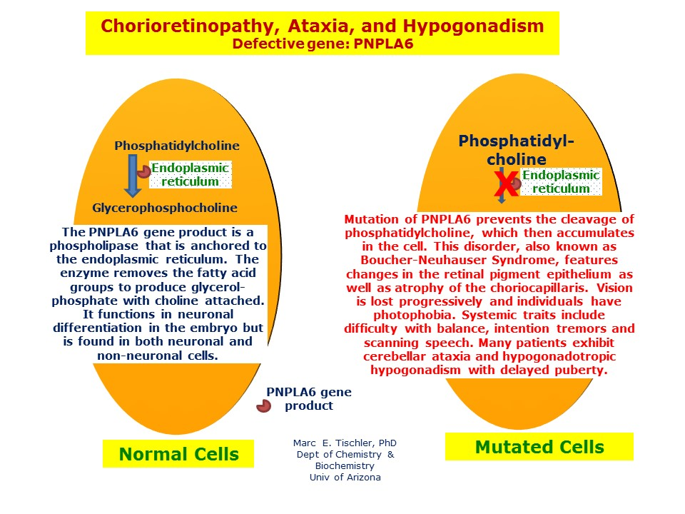
BNHS is an infrequent neurodegenerative disorder caused by mutations in the PNPLA6 gene on chromosome 19, inherited in an autosomal recessive manner. Both parents must carry one copy of the mutated gene for a child to be affected. These mutations disrupt neuronal and hormonal functions, leading to the syndrome's distinctive symptoms. With only about 22 documented cases, BNHS is part of a broader spectrum of PNPLA6-related disorders. Genetic testing is crucial for accurate diagnosis and effective management, allowing for informed genetic counseling and understanding of the inheritance complexities.
Differential Diagnosis
Clinicians should consider other similar conditions during diagnosis to rule out overlapping disorders that present with similar symptoms, such as cerebellar ataxia-hypogonadism syndrome.
Hormonal Evaluations
Blood tests are often conducted to measure hormone levels, including luteinizing hormone (LH), follicle-stimulating hormone (FSH), and testosterone, to assess for hypogonadotropic hypogonadism.
Conclusion
Ataxia-hypogonadism-choroidal dystrophy syndrome (BNHS) represents a unique intersection of neurodegenerative, hormonal, and visual impairments. Genetic research and clinical care advancements are paving the way for improved understanding and management despite its rarity. Raising awareness and fostering a supportive community can significantly enhance the quality of life for those affected. Ongoing research and patient advocacy are crucial in improving outcomes and offering hope to individuals and families affected by this complex disorder. Engaging with specialized medical professionals and patient organizations is vital for effective management. Increased awareness and support for BNHS can drive future research, unveiling novel therapeutic strategies and fostering a better quality of life for those impacted.
X-linked cerebral adrenoleukodystrophy (X-ALD): Mutation by ABCD1 gene
Introduction
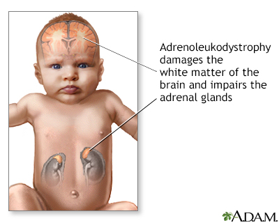
X-linked adrenoleukodystrophy (X-ALD) is the most common peroxisomal disorder. The disease is caused by mutations in the ABCD1 gene that encodes the peroxisomal membrane protein ALDP which is involved in the transmembrane transport of very long-chain fatty acids (VLCFA; ≥C22).They most frequently present in childhood (childhood cerebral ALD). However never before the age of 2.5 years. The childhood cerebral form of X-linked adrenoleukodystrophy typically occurs in boys. Girls are rarely affected with this type. In addition, damage to the outer layer of the adrenal glands (adrenal cortex) causes a shortage of certain hormones (adrenocortical insufficiency). Adrenocortical insufficiency may cause weakness, weight loss, skin changes, vomiting, and coma.
GENE INVOLVED
Variants (also known as mutations) in the ABCD1 gene cause X-linked adrenoleukodystrophy. The ABCD1 gene provides instructions for producing the adrenoleukodystrophy protein (ALDP), which is involved in transporting certain fat molecules called very long-chain fatty acids (VLCFAs) into peroxisomes. ABCD1 gene variants result in a shortage (deficiency) of ALDP. When this protein is lacking, the transport and subsequent breakdown of VLCFAs is disrupted, causing abnormally high levels of these fats in the body. The accumulation of VLCFAs may be toxic to the adrenal cortex and myelin.
PREVALENCE
The prevalence of X-linked adrenoleukodystrophy is 1 in 15,000 individuals worldwide. This condition occurs with a similar frequency in all populations.
FUTURE SCOPE
For the majority of patients with X-ALD there is currently no curative or preventive treatment. However, several promising new approaches will hopefully come to fruition in the future. For example, it has been demonstrated in X-ALD cells that small interfering RNA (siRNA)-mediated inhibition of ELOVL1 reduces VLCFA synthesis and level.
Sturge–Weber Syndrome(SWS) - Port-Wine Stains Caused by Somatic Mutation in GNAQ
Introduction
Sturge-Weber syndrome (SWS) is a rare neurocutaneous syndrome characterized by angiomas involving the face, choroid, and leptomeninges.The neurologic manifestations of SWS include atonic, tonic, or myoclonic seizures.Sturge-Weber syndrome is a condition that affects the development of certain blood vessels, causing abnormalities in the brain, skin, and eyes from birth. Sturge-Weber syndrome has three major features: a red or pink birthmark called a port-wine birthmark, a brain abnormality called a leptomeningeal angioma, and increased pressure in the eye (glaucoma).
GENE INVOLVED
Sturge-Weber syndrome is a sporadic developmental disorder caused by somatic mosaic mutations in the GNAQ gene, which is located on the long arm of chromosome 9. The presence of facial and pial angiomas is thought to result from the persistence of primordial sinusoidal vascular channels. Another hypothesis suggests that incomplete development of the brain's superficial venous drainage system leads to dilation of capillaries and small venous channels as a compensatory mechanism.
SIGN & SYMPTOMS
Most people with Sturge-Weber syndrome are born with a port-wine birthmark. This type of birthmark is caused by enlargement (dilatation) of small blood vessels (capillaries) near the surface of the skin. Port-wine birthmarks are typically initially flat and can vary in color from pale pink to deep purple. In people with Sturge-Weber syndrome, the port-wine birthmark is most often on the face, typically on the forehead, temple, or eyelid. The port-wine birthmark is usually only on one side of the face but can be on both sides. Over time, the skin within the port-wine birthmark can darken and thicken.
PREVALENCE
The incidence of Sturge-Weber syndrome is not well known and estimated to be 1 in 20,000-50,000 live births. SWS affects males and females equally and there is no race predilection
FUTURE SCOPE
Early diagnosis of SWS brain involvement and prediction of seizure onset and other neurologic outcomes is becoming increasingly urgent. As effective treatment approaches are developed, it is likely that new interventions, possibly able to delay or prevent seizure onset and thus decrease cognitive deterioration, will be considered. Small trials should continue, and when results are promising, multicenter efforts must be implemented to further assess these approaches
Spinocerebellar ataxia type 6: Calcium channel genetics with involvement of CACNA1A
Introduction
Spinocerebellar ataxia type 6 (SCA6) is a neurodegenerative disorder marked by a gradual decline in motor function. Spinocerebellar ataxia type 6 (SCA6) is caused by small expansions of a CAG repeat in the 3′ region of the CACNA1A gene, which encodes the α1A subunit of P/Q-type voltage-gated calcium channels. These channels are primarily expressed in cerebellar Purkinje and granule cells. SCA6 is one of three disorders linked to mutations in the CACNA1A gene, alongside episodic ataxia type 2 (EA2), typically caused by protein-truncating mutations, and familial hemiplegic migraine, which arises from missense mutations. While EA2 and familial hemiplegic migraine are considered channelopathies due to alterations in P/Q channel function, SCA6 involves CAG repeat expansions, which may lead to a toxic gain-of-function mechanism similar to other polyglutamine repeat disorders.
PREVALENCE
The worldwide prevalence of SCA6 is estimated to be less than 1 in 100,000 individuals.
SIGN & SYMPTOMS
Early symptoms commonly include difficulties with coordination and balance (ataxia), as well as speech impairments (dysarthria), involuntary eye movements (nystagmus), and double vision. As the condition progresses, individuals may experience worsening coordination in their arms, tremors, and involuntary muscle contractions (dystonia). Typically, symptoms emerge in a person’s forties or fifties, and by their sixties, most affected individuals require a wheelchair for mobility assistance.
GENE INVOLVEMENT
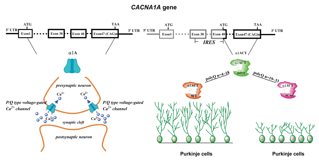
Spinocerebellar ataxia type 6 (SCA6) is the prototype of a pure cerebellar ataxia, associated with a severe form of progressive ataxia and cerebellar dysfunction. SCA6 is caused by a CAG repeat expansion in the CACNA1A gene which encodes the α1A subunit of the P/Q-type voltage-gated calcium channel.
FIG : Involvement of CACNA1A gene in spinocerebellar ataxia type 6
FUTURE SCOPE
Further complicating our understanding of this channel's function and regulation is the fact that several splicing variants of CACNA1A exist, and the GAG expansion is only predicted to be present in some of these isoforms. How this differential mRNA splicing might contribute to phenotypic variability and why some patients have episodic features is unknown. Structure-function studies in simplified systems or transgenic mice are necessary to clarify these issues.
Pitt–Hopkins syndrome: Due to loss of TCF4-regulated gene transcription
Introduction
Pitt-Hopkins syndrome (PTHS) is a rare genetic disorder that affects the nervous system. Children with this condition typically have unique facial features, intellectual disabilities, delayed developmental milestones, and difficulties with speech. They may also experience seizures, abnormal breathing patterns, poor coordination (ataxia), and repetitive hand movements. Other common issues include constipation, sleep problems, and severe nearsightedness (myopia). While behavioral challenges are frequent, affected children are often described as sociable and cheerful. Some may also meet the criteria for autism spectrum disorder. The severity and specific symptoms of PTHS can vary between individuals. This condition is caused by mutations in the TCF4 gene.
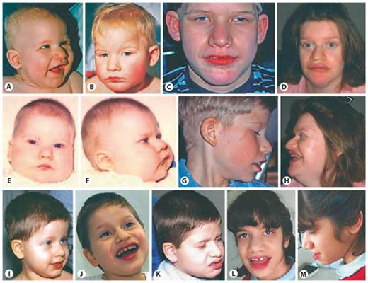
FIG: Facial features of patients with TCF4-related Pitt-Hopkins syndrome
PREVALENCE
Pitt-Hopkins syndrome is thought to be a very rare condition. Approximately 500 affected individuals have been reported worldwide
GENE INVOLVEMENT
Mutations in the TCF4 gene lead to Pitt-Hopkins syndrome. The TCF4 gene encodes a protein that binds to other proteins and specific regions of DNA, helping regulate the activity of numerous other genes. Due to its role in binding DNA and controlling gene expression, TCF4 is classified as a transcription factor. This protein is important for cell differentiation, where cells mature to perform specific functions, and it also plays a role in the process of programmed cell death (apoptosis).
SIGN AND SYMPTOMS
Individuals with Pitt-Hopkins syndrome often experience learning disabilities and developmental delays, such as delayed milestones like walking and lack of speech. They may also have distinctive facial features and exhibit changes in breathing patterns, including episodes of hyperventilation or apnoea, where breathing briefly stops before resuming. Additionally, those with Pitt-Hopkins may suffer from seizures (epilepsy) and digestive issues, particularly constipation.
FUTURE SCOPE
As a future development, it could be tested on images of patients with other dysmorphic syndromes, especially those requiring differentiation from PTHS. In conclusion, it is possible that the coupling of the phenotype analysis, done by computer vision algorithms, with the continuously growing genomic knowledge, will open new ways to rapidly reach an accurate molecular diagnosis for patients with genetic syndromes, and may become a key-factor in the field of precision medicine. In future studies, it could be possible to combine the facial analysis described here with genome sequencing data. This will enable improved prioritization of gene-variant results.
Short stature-pituitary and cerebellar defects-small sella turcica syndrome : A mutation in the LHX4 gene
Introduction
Short stature-pituitary and cerebellar defects-small sella turcica syndrome is a rare genetic disorder marked by short stature, anterior pituitary hormone deficiencies, a small sella turcica (the bony structure housing the pituitary gland), and an underdeveloped anterior pituitary. The condition is also characterized by pointed cerebellar tonsils and, in some cases, posterior pituitary ectopia (misplaced posterior pituitary). The syndrome has been observed across three generations in a large French family and follows an autosomal dominant inheritance pattern. The underlying cause is a germline mutation in the LIM-homeobox transcription factor LHX4 gene, located at 1q25.
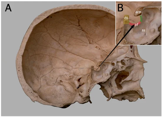
FIG: (A) General view. (B) Enlargement of the sella turcica, showing its boundaries. The anterior wall of the sella is marked pale green. The pituitary fossa (pf) on the sella floor is marked pale pink.
SYMPTOMS
The syndrome presents with short stature, a small sella turcica, hypoplasia of the anterior pituitary gland, and pointed cerebellar tonsils. Some individuals may also have ectopia of the posterior pituitary gland.
GENE INVOLVEMENT
A mutation in the LHX4 gene disrupts the production of functional proteins that play a key role in pituitary development. These abnormal proteins exhibit reduced DNA-binding ability, specifically impairing their interaction with the PIT1 promoter, which is crucial for normal pituitary hormone regulation and function.
PREVALENCE
This rare syndrome, with an occurrence of less than 1 in 1,000,000, follows an autosomal dominant inheritance pattern and typically manifests in infancy or the neonatal period.
FUTURE SCOPE
The future scope for research and clinical management of this syndrome includes:
Genetic Studies: Understanding the genetic basis of short stature and associated defects could lead to better diagnostic tools and targeted therapies.
Longitudinal Studies: Tracking growth patterns and hormonal changes over time in affected individuals will help refine treatment protocols.
Hormonal Therapies: Development of more effective hormone replacement therapies tailored to individual needs could enhance quality of life.
Multidisciplinary Approaches: Integrating endocrinology, neurology, and genetics in treatment plans may provide comprehensive care for patients.
SETBP1 hotspot results in a Schinzel–Giedion syndrome
Introduction
Schinzel-Giedion syndrome (SGS), an ultra-rare multisystem disorder caused by gain-of-function pathogenic variants in a SETBP1 mutational hot spot.It is characterized by global neurodevelopmental impairment leading to moderate-to-profound intellectual disability, epilepsy (often refractory to treatment), hypotonia, spasticity, dysautonomia, hearing loss, and cerebral visual impairment.
SIGN & SYMPTOMS
Symptoms include poor weight gain often associated with gastroesophageal reflux disease, chronic vomiting, constipation, gastroparesis, and/or feeding intolerance. Structural malformations can involve the heart, skeleton, kidney and urinary tract, genitalia, and brain. Anomalies of the liver, spleen, and/or pancreas are less common. Other features may include neuroepithelial neoplasia, severely disrupted sleep, choanal stenosis, inguinal hernia, sensitive skin, and increased risk of infection.
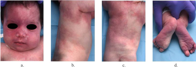
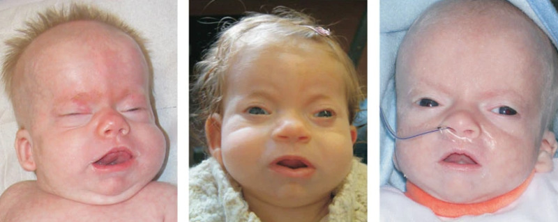
FIG: Characteristic facial features in individuals with Schinzel-Giedion syndrome (SGS)
PREVALENCE
To date, over 50 individuals with molecularly confirmed classic Schinzel-Giedion syndrome (SGS) have been reported in the medical literature. Additionally, more than 40 individuals were clinically diagnosed with classic SGS before SETBP1 pathogenic variants were identified as the causative factor.
GENE INVOLVEMENT
Schinzel-Giedion syndrome (SGS) is caused by pathogenic heterozygous gain-of-function variants within a mutational hot spot of SETBP. This mutational hot spot is comprised of a 12-base-pair region within exon 4 encoding for amino acid residues 868 to 871 within the SKI domain.
Progressive encephalopathy with leukodystrophy due to DECR deficiency is a rare mitochondrial disease - Results from mutations in the NADK2 gene
Introduction
Progressive encephalopathy with leukodystrophy due to DECR deficiency is a rare mitochondrial disease, which presents with neonatal hypotonia, central nervous system abnormalities (ventriculomegaly, corpus callosum hypoplasia, cerebellar atrophy), acquired microcephaly, failure to thrive Progressive, developmental delay and intermittent lactic acidosis provoked by catabolic stress (e.g. infection). Hyperlysinemia and elevated C10:2 carnitine can be detected in plasma. Later on, epilepsy, cerebellar ataxia, renal tubular acidosis, severe encephalopathy, dystonia, spastic quadriplegia and other complications may develop.
GENE INVOLVEMENT
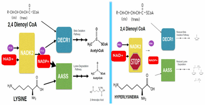
This disorder results from mutations in the NADK2 gene, which encodes mitochondrial NAD kinase. NAD kinase is critical for the production of NADPH, a cofactor required by DECR (2,4-dienoyl-CoA reductase), a mitochondrial oxidoreductase enzyme. The deficiency of NADPH impairs DECR activity, leading to mitochondrial dysfunction and the accumulation of toxic metabolic byproducts.
FIGURE: Model of metabolic dysfunction in NADK2 deficiency (right) compared to normal (left)
SYMPTOMS
The condition is characterized by hypotonia, central nervous system abnormalities, microcephaly, failure to thrive, developmental delay, and lactic acidosis. As the disease progresses, patients may develop complications such as epilepsy, cerebellar ataxia, renal tubular acidosis, severe encephalopathy, dystonia, and spastic quadriplegia.
PREVALENCE
Occurrence in 1 / 1 000 000,Inheritance- Autosomal recessive therefore age of onset: Infancy, Neonatal
FUTURE SCOPE
For future research, we would suggest performing prospective follow-up of these patients, for example with the Newcastle Paediatric Mitochondrial Disease Scale. Only a prospective follow-up will provide valid data to be used in the preparation of clinical trials in predicting the natural disease course or selecting relevant outcome measures.
We also suggest continuing publishing clinical details of these rare diseases. Only more case descriptions will enable us to predict the natural disease course of patients with these rare diseases, which is not only useful for future clinical trials, but is also indispensible for the patients and their families.