Small interfering RNA (siRNA) a molecular tool in the therapeutic landscape for
Duchenne muscular dystrophy (DMD). By targeting specific mRNAs, siRNAs can
modulate gene expression and potentially restore dystrophin production or its
functional substitutes, thereby addressing the root cause of the disease.
1. siRNA Mechanism
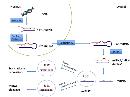
siRNA works by incorporating into the RNA-induced silencing complex (RISC),
leading to the degradation of complementary mRNA strands. This pathway inhibits
the expression of genes involved in the progression of DMD, enabling the possibility
to silence mutated genes or upregulate compensatory proteins necessary for muscle
function enhancement.
2. Targeting Dystrophin Mutations
In DMD, siRNA can specifically target the mutations in the dystrophin gene, aiming
to reduce the production of mutated dystrophin. This is crucial as the absence of
functional dystrophin leads to muscle degeneration. By deploying siRNA therapeutics,
researchers intend to decrease the expression of flawed transcripts while promoting
the synthesis of functional dystrophin or related Protein.
3. Potential in Alternative Splicing
siRNA also facilitates alternative splicing strategies, which can accurately modify
Dystrophin’s mRNA to better suit therapeutic needs. For instance, siRNA can help
reframe exons that are incorrectly spliced, potentially leading to the production of a
truncated but functional form of dystrophin.
4. Delivery Challenges
The successful therapeutic application of siRNA in DMD faces challenges,
particularly in the effective delivery of siRNA molecules to muscle tissues. Innovative
vector systems, such as adeno-associated viruses (AAVs), are being explored to
enhance the in vivo delivery of siRNA to target affected muscle tissues.
5. Clinical Perspectives
Research on siRNA as a therapeutic strategy for DMD is ongoing, with early
preclinical studies indicating potential efficacy in restoring dystrophin levels and
promoting muscle regeneration. However, further clinical trials are essential to
validate these findings and assess the long-term effects and safety of siRNA therapies
for DMD.
In summary, siRNA holds significant promise as a therapeutic strategy for
Duchenne muscular dystrophy, primarily through its ability to manipulate gene
expression and target the underlying genetic mutations associated with the disease.
Further researches will clarify its efficacy and viability in clinical settings.
DMD is primarily associated with mutations in the dystrophin gene, studies have shown that RYR1 dysfunction also plays a vital role. In DMD, abnormal RYR1 function leads to impaired calcium ion handling in muscle cells, which contributes to delayed muscle differentiation and disease progression. Stabilizing compounds that target RYR1, such as Rycals, have shown promise in improvement of muscle function in preclinical models, suggesting that RYR1 could be a therapeutic target in DMD(MDPI).
These studies highlight the critical role of RYR1 in muscle function and its potential as a target for therapeutic interventions in various muscular dystrophies and related myopathies2
Rycals are a class of small molecules designed to stabilize the ryanodine receptor 1 (RYR1) calcium channel in muscle cells, preventing abnormal calcium leakage from the sarcoplasmic reticulum, which is critical for proper muscle function.
Mode of Action:
1. Stabilization of the RYR1-Calstabin Complex:
RYR1 channels are responsible for releasing calcium ions from the sarcoplasmic reticulum into the cytoplasm, a crucial step in muscle contraction. These channels are regulated by a protein called calstabin1 (FKBP12), which binds to RYR1 and helps maintain the channel in a closed state when it is not supposed to be active.
In conditions like muscular dystrophy, oxidative stress and other pathological signals cause post-translational modifications of RYR1, such as S-nitrosylation or phosphorylation. These modifications can disrupt the binding of calstabin1 to RYR1, leading to a "leaky" channel that allows calcium to escape continuously into the cytoplasm, even when it should be closed.
2.Preventing Calcium Leak:
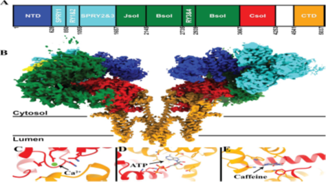
Fig. A Schematic diagram of the domain architecture of RyR1. B Coulombic density map of RyR1 (PDBID: 7M6A).The accessory protein calstabin-1 is shown in yellow. showing Calcium (C), ATP (D), and caffeine (E) binding sites with the coordinating residues
Rycals stabilize the interaction between RYR1 and calstabin1, preventing the dissociation of calstabin1 even under conditions that would normally cause it to detach. By doing so, Rycals help to keep the RYR1 channels closed when they are not supposed to release calcium, thereby reducing calcium leakage.
3.Restoring Calcium Homeostasis:
By preventing the abnormal calcium leak, Rycals help restore proper calcium homeostasis within the muscle cells. This is crucial because excessive cytoplasmic calcium can lead to muscle damage, impaired muscle contraction, and eventually contribute to muscle weakness and degeneration, as seen in various muscular dystrophies.
4.Therapeutic Implications:
Preclinical studies, particularly in animal models of muscular dystrophy, have shown that treatment with Rycals can reduce muscle damage, improve muscle function, and delay the progression of the disease. These compounds are being explored as potential therapeutic agents for conditions where RYR1 dysfunction is a key contributor to pathology, including certain types of muscular dystrophy and heart diseases.
In summary, Rycals act by stabilizing the RYR1-calstabin1 complex, preventing pathological calcium leakage from muscle cells, thereby maintaining muscle function and reducing damage in muscular dystrophies
LAMA2 congenital muscular dystrophy (LAMA2-CMD) is a serious neuromuscular disorder caused by mutations in the LAMA2 gene, leading to a deficiency in the laminin-α2 protein. It is the most common congenital muscular dystrophy and is characterized by severe muscle weakness, typically observed from early infancy. The disease can manifest with various clinical features, including hypotonia, muscle weakness, and structural brain abnormalities.Its prevalence varies globally and research continues to explore potential therapeutic interventions.
1. Definition
LAMA2 congenital muscular dystrophy (LAMA2-CMD) is the most prevalent form of
congenital muscular dystrophy, often resulting in severe outcomes for affected individuals.The disease arises from mutations in the LAMA2 gene, which encodes the laminin-α2 chain,an essential component of the extracellular matrix in muscle tissues. This deficiency can impair muscle development and function, leading to serious mobility issues.
2. Clinical Features
Patients with LAMA2-CMD typically present with peripheral hypotonia and muscle
weakness from an early age, often leading to delayed motor milestones. The condition may also be associated with cognitive impairments and neurological abnormalities observable on imaging studies, indicating broader involvement beyond just muscle.
3. Prevalence and Epidemiology
The estimated prevalence of LAMA2-CMD ranges significantly, with reports indicating
occurrences between 1 to 9 per 1,000,000 children. Genetic assessments indicate that
recessive mutations in the LAMA2 gene are prevalent in many populations, which informs strategies for diagnosis and counselling for affected families.
4. Pathophysiology
The underlying pathology of LAMA2-CMD is primarily linked to the absence or truncation
of the laminin-α2 protein, leading to disruptions in cellular adhesion and signalling in muscle tissues. This results in muscle fibre degeneration and fatty replacement, contributing to the progressive nature of the disease.
5. Research and Therapeutic Approaches
Current research is focusing on understanding the disease mechanisms more
comprehensively, with studies investigating potential treatments that target underlying
genetic mutations. Notable approaches include gene therapy, small molecule interventions,and enhanced supportive care strategies to manage symptoms and improve quality of life for affected individuals.
In conclusion, LAMA2 congenital muscular dystrophy poses significant challenges,
highlighting the need for continued research and therapeutic advancement
Hope on the Horizon: Innovative Approaches to Treating Autosomal Recessive Centronuclear Myopathy
Autosomal Recessive Centronuclear Myopathy (AR-CNM) is a rare genetic disorder that primarily affects skeletal muscle function. Characterized by muscle weakness that can range from mild to severe, AR-CNM is caused by mutations in specific genes involved in muscle fiber maintenance and organization. Unlike other forms of centronuclear myopathy (CNM), AR-CNM is inherited in an autosomal recessive pattern, meaning both copies of a faulty gene (one from each parent) are required for the disease to manifest.
Genetic Basis of AR-CNM
AR-CNM is most commonly associated with mutations in the BIN1, RYR1, and TTN genes. These genes play critical roles in the structure and function of muscle cells, particularly in the organization of the sarcomere (the basic contractile unit of muscle). In affected individuals, muscle fibers often have nuclei located centrally rather than at the periphery, which is a hallmark of centronuclear myopathies. This abnormality disrupts normal muscle contraction and leads to progressive muscle weakness.
BIN1: Plays a role in membrane curvature and T-tubule formation, crucial for muscle excitation and contraction.
RYR1: Encodes the ryanodine receptor, which is involved in calcium release from the sarcoplasmic reticulum, essential for muscle contraction.
TTN: Encodes titin, a massive protein that acts as a molecular spring, helping muscles to stretch and recoil.
Symptoms and Clinical Presentation
The clinical spectrum of AR-CNM is broad, with some patients showing symptoms at birth (congenital myopathy) and others developing them later in childhood or adulthood. Common symptoms include:
Muscle Weakness: Affects proximal (near the body) and sometimes distal (far from the body) muscles.
Delayed Motor Milestones: Infants may take longer to sit up, crawl, or walk.
Respiratory Issues: In severe cases, respiratory muscles may be affected, leading to breathing difficulties that require mechanical ventilation.
Facial Muscle Weakness: This can result in difficulties with facial expressions, speech, and swallowing.
Skeletal Deformities: Some patients may develop scoliosis (curvature of the spine) or other orthopedic issues due to muscle weakness.
Diagnosis and Genetic Testing
Diagnosis of AR-CNM is based on a combination of clinical symptoms, muscle biopsy, and genetic testing. A muscle biopsy typically reveals the characteristic presence of centrally located nuclei in muscle fibers. However, genetic testing is the definitive diagnostic tool, identifying mutations in BIN1, RYR1, TTN, or other genes associated with centronuclear myopathies.
With advances in genetic screening techniques such as next-generation sequencing (NGS), identifying the specific mutation responsible for AR-CNM has become more accessible and efficient. This is crucial not only for diagnosis but also for determining the best course of treatment, which may be tailored to the specific mutation involved.
Current Treatments and Challenges
As of now, there is no cure for AR-CNM, and treatment is primarily supportive. Physical therapy, respiratory support, and orthopedic interventions can improve quality of life, but these approaches do not address the underlying genetic cause.
Antisense Oligonucleotide (ASO) Therapy: A Promising Avenue
Recent advances in gene therapy, particularly antisense oligonucleotide (ASO) technology, offer hope for patients with AR-CNM. ASOs are short, synthetic strands of nucleotides designed to target and modify specific RNA sequences, potentially correcting the expression of mutated genes at the transcriptional level. This precision-targeting approach allows for the selective silencing, splicing correction, or degradation of faulty RNA transcripts, which could mitigate the effects of the disease-causing mutation.
For example, in certain cases of AR-CNM, ASOs could be designed to:
Modulate Splicing: Correct aberrant splicing caused by mutations in genes like BIN1 or RYR1, restoring normal protein function.
Reduce Toxic RNA/Proteins: Suppress the expression of defective proteins that cause muscle degeneration.
Compensate for Gene Loss: Increase the expression of healthy genes to compensate for loss-of-function mutations.
While ASO therapy is still in the experimental stages for AR-CNM, early studies in similar neuromuscular disorders (such as spinal muscular atrophy and Duchenne muscular dystrophy) have shown promise. These successes suggest that ASO technology could play a transformative role in AR-CNM treatment.
Research and Development in AR-CNM
Research into the pathophysiology and genetics of AR-CNM is ongoing, with a growing emphasis on developing molecular therapies like ASOs. In addition, patient registries and natural history studies are critical for understanding the progression of the disease and identifying biomarkers that could guide therapeutic development. Collaborative research efforts involving scientists, clinicians, and patient advocacy groups are accelerating progress in this area.
Our company is actively engaged in the development of antisense oligonucleotide therapies for rare neuromuscular disorders like AR-CNM. By focusing on precision medicine approaches, we aim to provide targeted treatments that address the underlying genetic causes of these diseases. The future of AR-CNM treatment lies in personalized medicine, where therapies are designed based on each patient's unique genetic makeup.
Support for Patients and Families
For patients living with AR-CNM, access to multidisciplinary care is essential. This includes physical therapy to maintain muscle function, respiratory support, and orthopedic care. Additionally, patient advocacy groups and online communities provide crucial emotional and practical support for patients and families navigating the complexities of living with a rare disease.
Organizations dedicated to rare diseases, such as the Muscular Dystrophy Association (MDA) and the Myotubular Trust, offer resources and support networks. These organizations often facilitate connections between families, researchers, and clinicians, fostering a community dedicated to advancing research and improving patient outcomes.
Conclusion
Autosomal Recessive Centronuclear Myopathy is a rare but debilitating disorder that significantly impacts patients' quality of life. However, with advances in genetic research and therapies like antisense oligonucleotides, there is growing hope for more effective treatments in the future. Our company is at the forefront of this exciting field, committed to developing innovative therapies that could change the landscape of AR-CNM treatment and improve the lives of those affected by this rare condition.
By harnessing the power of ASO technology, we believe that precision medicine offers the best path forward for addressing the unmet medical needs of patients with AR-CNM and other rare genetic disorders. Stay tuned as we continue to make strides in our research and clinical development programs, bringing us one step closer to delivering life-changing treatments.
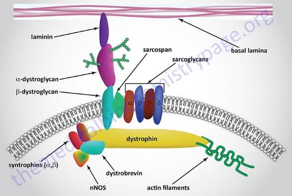
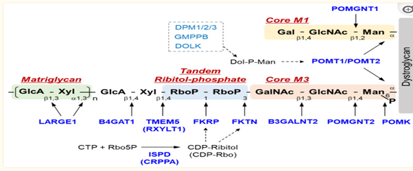
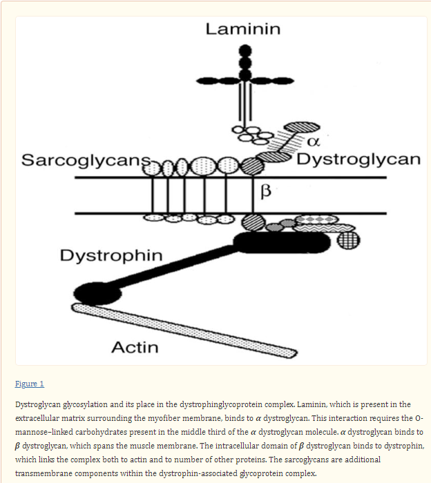
The LARGE1 gene encodes a glycosyltransferase, which critically contributes to the post-translational modification of alpha-dystroglycan (α-DG), a transmembrane receptor fundamental in anchoring the extracellular matrix to the cytoskeleton of muscle cells. This glycosylation process is essential for the proper functioning of α-DG, ensuring the stability and functionality of muscle tissues.
Mutations and Path of mechanism
Mutations in the LARGE1 gene disrupt the normal glycosylation of α-DG, leading to congenital muscular dystrophy type 1D (MDC1D). MDC1D is characterized by severe muscle weakness, brain abnormalities, and intellectual disability. The mutation prevents the proper glycosylation of α-DG, which is critical for its role in muscle cell integrity.
Clinical Features
MDC1D resulting from LARGE1 gene mutations manifests in severe mental retardation and abnormal glycosylation of α-DG. Patients typically present with congenital muscular dystrophy features, including pronounced muscle weakness and periodic electroretinogram anomalies. Brain abnormalities and intellectual disabilities are prominent clinical features.
Molecular Impact
The primary consequence of mutations in the LARGE1 gene is defective glycosylation of α-DG. This hypoglycosylation reduces the ligand-binding capacity of α-DG, impairing its anchoring function between the extracellular matrix and the muscle cell cytoskeleton. Research on animal models, specifically the Large myd mouse, demonstrates that α-DG from these models is hypoglycosylated and exhibits reduced ligand-binding activities akin to human dystroglycanopathies.
Fig. Above diagram showing LARGE1 & other genes mode of action in muscle tissues.
Potential Therapeutic Approaches
Gene therapy targeting the LARGE1 gene holds promise in treating MDC1D. Techniques such as adenoviral-mediated gene transfer have been successful in restoring muscle function and prolonging survival in preclinical models. Overexpression of LARGE1 in animal models has been shown to induce hyper glycosylation of α-DG, compensating for defective glycosylation pathways in various glycosyltransferase-deficient muscular dystrophies.
Future Research Directions
Ongoing research is focusing on optimizing gene therapy protocols for clinical application. Studies are exploring the use of adeno-associated viral (AAV) vectors to efficiently deliver the LARGE1 gene into muscle tissues, aiming for sustained expression and minimal side effects. There is also an interest in developing therapies that aim to restore full-length dystrophin in patients with Duchenne muscular dystrophy (DMD) by using multi-vector systems to deliver the entire gene.
Conclusion
The LARGE1 gene plays an integral role in the glycosylation of α-DG, and its mutations lead to congenital muscular dystrophy type 1D (MDC1D). This results in significant muscle weakness, brain abnormalities and intellectual disabilities. Gene therapy targeting LARGE1 shows considerable promise for treating MDC1D by correcting the defective glycosylation of α-DG, showing a potential avenue for effective treatments in the future.
Primary mitochondrial myopathies (PMM) represent a group of genetic disorders characterized by dysfunction in mitochondrial energy production, predominantly impacting skeletal muscle. These conditions manifest through a variety of symptoms influenced by the underlying genetic mutations affecting mitochondrial DNA or nuclear DNA.
1. Definition and Overview
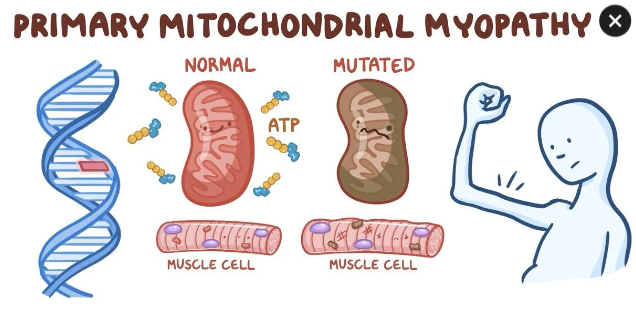
Primary mitochondrial myopathies (PMM) are genetically defined disorders leading to defects in oxidative phosphorylation, thereby affecting predominantly skeletal muscle. These disorders are associated with variations in the genetic material found in mitochondrial DNA (mtDNA) or nuclear DNA, impairing the cells' ability to produce energy efficiently. Mitochondria play a crucial role in energy metabolism by generating adenosine triphosphate (ATP), the primary energy carrier in cells. This energy deficit can significantly impact high-energy tissues such as muscles, brain, and heart.
2. Symptoms and Variability
The signs and symptoms of PMM vary widely among individuals, even those sharing the same genetic mutations. Common symptoms include muscle weakness, fatigue, and specific muscle diseases, such as mitochondrial myopathy. Progressive external ophthalmoplegia, characterized by paralysis of the eye muscles, is one of the most frequent presentations.
3. Genetic Basis and Inheritance
PMM can arise from mutations in genes that provide essential instructions for mitochondrial function. Genetic variations can be inherited in different patterns, including autosomal recessive and dominant forms, or can occur spontaneously. Approximately half of mitochondrial proteins are encoded by nuclear genes, while the remaining are derived from mtDNA, which is inherited maternally. The concept of heteroplasmy, where both mutated and normal mtDNA coexist, is also critical in understanding the diverse expression of symptoms within families.
4. Diagnosis
The diagnosis of PMM involves a detailed patient and family history, comprehensive clinical evaluation, and various specialized tests. Characteristic symptoms may overlap with other disorders, requiring a multidisciplinary diagnostic approach that could include biochemical tests, genetic analysis, and muscle biopsies. Early and accurate diagnosis is essential for effective management and treatment planning.
5. Treatment Options
Currently, there is no cure or disease-modifying treatment for PMM, and management strategies focus on alleviating symptoms and improving quality of life. Supportive care often includes a multidisciplinary approach involving various specialists, including neurologists, cardiologists, and physical therapists. Some patients may benefit from dietary supplements known as "mito-cocktails," although their efficacy can vary. Exercise programs have also been shown to improve strength and reduce fatigue in affected individuals.
6. Research and Future Directions
Emerging therapies, including gene therapy, hold promise for treating mitochondrial disorders by potentially correcting genetic defects. Current clinical trials are exploring innovative interventions, such as elamipretide, aimed at enhancing mitochondrial function. Continued research is crucial to better understand PMM and develop effective treatment protocols, ultimately improving outcomes for affected individuals.
7. Epidemiology
Mitochondrial diseases, including PMM, are among the most prevalent forms of metabolic diseases, with estimates suggesting they affect approximately 1 in 5,000 individuals in the general U.S. population. However, challenges in diagnosis and variability in symptoms can lead to underreporting, making it difficult to ascertain their true prevalence.
Autosomal dominant centronuclear myopathy (AD-CNM) is a type of centronuclear myopathy, which is a group of rare, inherited conditions that affect the muscles. Dynamin 2 (DNM2) plays a critical role in various aspects of neuromuscular diseases. Understanding its functions is essential for elucidating the pathophysiology of these conditions and may provide insights into potential therapeutic approaches.
1. Role in Membrane Dynamics
Dynamin 2 is a GTPase known for its function in membrane dynamics, particularly in endocytosis and the regulation of synaptic vesicles at the neuromuscular junction (NMJ). Through its ability to facilitate membrane fission, DNM2 is vital for the recycling of synaptic vesicles, which is crucial for proper neurotransmission.
2. Implications in Neuromuscular Disorders
Mutations in the DNM2 gene have been implicated in several neuromuscular disorders, including centronuclear myopathy and are associated with other congenital myopathies. These mutations disrupt the normal function of DNM2, leading to impaired muscle function and structural abnormalities at the NMJ.
3. Interaction with Other Proteins
DNM2 interacts with a variety of proteins involved in the assembly and maintenance of the NMJ, including elements crucial for synaptic adhesion and signaling. This interaction highlights its importance in maintaining NMJ integrity and facilitating effective neuromuscular communication.
4. Potential Therapeutic Targets
Research into the role of DNM2 in the context of neuromuscular diseases may lead to the identification of new therapeutic targets. By modulating DNM2 function or correcting its mutations, it might be possible to develop treatments that can restore proper muscle function and mitigate the effects of these diseases.
5. Research and Future Directions
Future studies are essential for further elucidating the role of DNM2 in neuromuscular diseases, particularly in understanding the mechanisms by which it contributes to disease pathology. This research may open new avenues for therapeutic interventions and improve outcomes for affected individuals
"Understanding Myotonic Muscular Dystrophy: A Deep Dive into the Most Common Adult Muscular Dystrophy"
Myotonic muscular dystrophy (MMD) encompasses a range of genetic disorders, primarily myotonic dystrophy type 1 (DM1) and type 2 (DM2), characterized by progressive muscle weakness and other systemic complications. The complexity of these diseases necessitates a thorough understanding of their genetic underpinnings, clinical manifestations, and available therapeutic approaches.
1. Definition
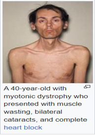
Myotonic dystrophy is the most common muscular dystrophy in adults, characterized by myotonia, muscle wasting and weakness, and multi systemic dysfunction. It is primarily categorized into two types: DM1 and DM2, both of which are inherited in an autosomal dominant manner.
2. Genetic Basis
DM1 is caused by unstable trinucleotide repeat expansions of CTG in the DMPK gene, while DM2 results from a (CCTG)n repeat expansion in the CNBP gene. These genetic alterations lead to variability in symptom presentation among patients, complicating diagnosis and treatment options.
3. Clinical Manifestations
The age of symptom onset for DM2 can range from 15 to 72 years, with proximal lower extremity weakness being the most common presenting symptom. Clinical features include myotonia, weakness, and, in some cases, cardiac abnormalities, which may lead to severe complications including heart failure
4. Diagnosis and Assessment
Diagnosis often involves genetic testing and clinical evaluation. Electromyography is also a valuable tool, as it can reveal myotonic discharges in patients. Additionally, the duration from symptom onset to diagnosis averages around 7.4 years, highlighting the challenges in early detection.
5. Treatment Options
Treatment for myotonic dystrophy focuses on managing symptoms and preventing complications. Although there is ongoing research into targeted therapies such as antisense oligonucleotides, no definitive cure currently exists. Regular monitoring and supportive care are crucial for improving quality of life.
6. Quality of Life Impact
MMD significantly impacts patients' quality of life due to its chronic nature and progressive symptoms. Multidisciplinary approaches involving physical therapy and psychological support can help address the varied challenges faced by patients.
7. Research and Future Directions
Current research is directed towards understanding the molecular pathogenesis of MMD through omics studies, which may pave the way for novel therapeutic strategies. Advances in gene therapy and novel pharmacological agents are essential for improving patient outcomes in the future.
8. Conclusion
In summary, myotonic muscular dystrophy represents a complex set of neuromuscular disorders requiring comprehensive clinical management and ongoing research to provide better treatment pathways and improve patient care.
Unlocking the Potential of miR-486-5p: Hope for Muscular Dystrophy"
Downregulation in Duchenne Muscular Dystrophy (DMD)
MicroRNAs (miRNAs) are short non-coding RNAs, highly conserved between species, that are powerful regulators of gene expression. Aberrant expression of miRNAs alters biological processes and pathways linked to human disease. miR-486-5p is a muscle-enriched miRNA localized to the cytoplasm and nucleus, and is highly abundant in human plasma and enriched in small extracellular vesicles.
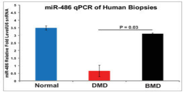
miR-486-5p expression is specifically downregulated in the skeletal muscle of patients with Duchenne Muscular Dystrophy (DMD), as compared to healthy controls. This downregulation has been observed consistently in both murine models and clinical samples from DMD patients, indicating its potential role in the disease pathology.
Biomarker for Disease Progression
miR-486-5p has been identified as a useful biomarker for dystrophic disease progression. Its expression levels correlate with muscle remodeling and the severity of the disease, making it a valuable candidate for monitoring the progression of muscular dystrophy and the effectiveness of therapeutic interventions.
Mechanistic Pathways
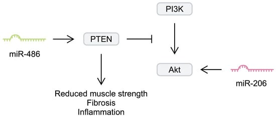
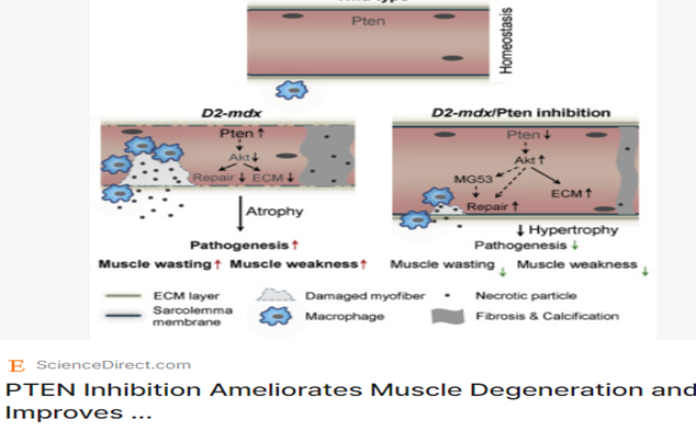
Overexpression of miR-486-5p has been shown to ameliorate disease progression in dystrophin-deficient skeletal muscle4. miR-486-5p induces skeletal muscle hypertrophy and ameliorates muscular dystrophy through the PTEN/AKT pathway. It directly targets PTEN,
Overexpression of miR-486-5p has been shown to ameliorate disease progression in dystrophin-deficient skeletal muscle4. miR-486-5p induces skeletal muscle hypertrophy and ameliorates muscular dystrophy through the PTEN/AKT pathway. It directly targets PTEN,
Potential Therapeutic Implications
Given its significant role in muscle remodeling and hypertrophy, miR-486-5p has therapeutic potential for DMD. In murine models, overexpression of miR-486-5p has led to improved muscle histology and prevention of myofiber degeneration69. Additionally, the transcriptional activation of miR-486-5p by the PAX3-FOXO1 transcription factor suggests that modulation of its expression could be a strategic therapeutic approach5.
Summary
miR-486-5p is a critical microRNA involved in the pathogenesis of Duchenne Muscular Dystrophy, primarily characterized by its downregulation in dystrophin-deficient muscle. It serves as a biomarker for disease progression and has potential therapeutic implications due to its role in modulating the PTEN/AKT signaling pathway and inducing muscle hypertrophy.
Overall, targeting miR-486-5p could represent a promising strategy for developing treatments aimed at alleviating the symptoms and progression of Duchenne Muscular Dystrophy.
"Gene Therapy Advances: How DYNE-101 Offers Hope for Myotonic Dystrophy-1"
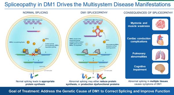
Myotonic dystrophy type 1 (DM1) is a rare neuromuscular disorder with multi systemic presentation. It is caused by expansion of CUG repeats in the 3’-UTR of the dystrophia myotonica protein kinase (DMPK) RNA which form hairpin-loop structures that sequester splicing regulators into toxic nuclear foci. It leads to widespread dysregulation of RNA splicing (spliceopathy) that drives the multisystem clinical manifestations.
DYNE-101 is an investigational therapeutic demonstrating significant potential in the treatment of myotonic dystrophy type 1 (DM1). It operates through targeted delivery of antisense oligonucleotides, aiming to correct splicing defects caused by toxic RNA accumulation. Early clinical trials indicate promising safety profiles and efficacy outcomes, including improvements in muscle function and splicing correction.
1. Mechanism of Action
DYNE-101 is designed to target nuclear DMPK RNA, the root cause of DM1 due to CUG repeat expansions. The drug consists of a gapmer antisense oligonucleotide (ASO) conjugated to a fragment antibody that binds to transferrin receptor 1 (TfR1)1. This targeting facilitates efficient delivery to muscle tissues, where it can promote degradation of the toxic RNA and restore normal splicing functions.
2. Clinical Trial Insights
The ACHIEVE trial, a Phase 1/2 study, evaluates the safety, tolerability, and efficacy of DYNE-101 in adults with DM1. Initial findings reported positive dose-dependent splicing correction among participants, particularly noted in the 5.4 mg/kg cohort, which exhibited a 27% mean correction after three months. The trial also suggests that all evaluable patients in this cohort experienced splicing correction across a broad 22-gene panel.
3. Functional Improvements
Patients treated with DYNE-101 have shown marked improvements in muscle function and strength, assessed through various clinical endpoints. Specifically, improvements were seen in tests like the 10-Meter Walk/Run test and the 5 Times Sit to Stand Test. Additionally, notable enhancements in the Myotonic Dystrophy Health Index (MDHI) were reported, indicating benefits in both physical ability and overall quality of life.
4. Safety Profile
The safety and tolerability of DYNE-101 have been favorable, with most treatment-emergent adverse events (TEAEs) classified as mild or moderate. Common reported side effects included nasopharyngitis and fatigue, with no participants demonstrating treatment-emergent anemia or serious adverse events directly related to the drug. Importantly, elevated liver enzymes were noted in some participants without affecting overall liver function.
5. Future Directions
Dyne Therapeutics continues to explore the full potential of DYNE-101, with further data expected in upcoming years. This ongoing research aims to solidify the drug's role in addressing the unmet medical needs of DM1 patients, with the hope of progressing towards accelerated regulatory pathways based on clinical efficacy and safety profiles.
"miR-486-5p and Its Therapeutic Potential in Spinal Muscular Atrophy"
Introduction
MicroRNAs (miRNAs) are a novel class of small non-coding RNAs that negatively regulate gene expression at the post-transcriptional level. miR-486-5p is a specific miRNA that has gained considerable attention due to its role in muscle differentiation and regeneration.
Role of miR-486-5p in Muscle Physiology
miR-486-5p is a muscle-enriched miRNA found in the cytoplasm and nucleus, and is highly abundant in human plasma and enriched in small extracellular vesicles. It plays an important role in the differentiation of satellite cells and myocytes by targeting the Paired box gene 3 (PAX3), PAX7, and MTSN4.
miR-486-5p and Spinal Muscular Atrophy
Spinal Muscular Atrophy (SMA) is a genetic disease characterized by the loss of motor neurons in the spinal cord, leading to muscle atrophy. Although specific studies directly linking miR-486-5p to SMA are sparse, miRNAs, including miR-486-5p, are understood to be crucial in various pathogenic events associated with SMA. Broadly, miR-486-5p’s involvement in muscle differentiation and regeneration positions it as a significant player in muscular atrophy conditions, such as SMA.
Mechanisms of miR-486-5p in Muscular Atrophy
miR-486-5p targets components of the Smad-dependent TGF-β signaling pathway, conferring protective effects in models of cardiac and pulmonary fibrosis. In the context of muscle biology, this signaling pathway is implicated in the regulation of muscle atrophy and hypertrophy, making miR-486-5p a potential modulator of muscular atrophy in SMA.
Regulatory Networks Involving miR-486-5p
Several studies have shown the regulatory roles miR-486-5p plays in muscle differentiation. It is known to repress the transcription of MRTF-A by binding to its 3′-untranslated region (UTR), which is pivotal for myoblast differentiation. Furthermore, modulation of miR-486-5p activity impacts satellite cell proliferation and differentiation, thereby influencing overall muscle homeostasis.
Implications in SMA Pathology
In SMA, motor neuron degeneration represents a primary pathological feature. miR-486-5p, by influencing muscle cell differentiation and growth, may affect the overall muscle integrity and response to motor neuron loss. Reduced levels of early motor neuron markers in SMA have been linked to alterations in miRNA expressions, including miR-486-5p7. Thus, miR-486-5p might play a part in SMA pathology by contributing to muscle degeneration and loss.
Potential Therapeutic Implications
Given its significant role in muscle physiology, miR-486-5p presents as a promising therapeutic target. Strategies aimed at modulating miR-486-5p levels could potentially ameliorate muscle atrophy in SMA. For instance, its downregulation has shown to improve hind limb functional recovery in mouse models of spinal cord injury (SCI), signifying its therapeutic potential in neuromuscular diseases.
Conclusion
miR-486-5p is integral in muscle differentiation, regeneration, and overall muscle homeostasis. While direct studies linking miR-486-5p to SMA are limited, the miRNA's established role in muscular physiology and its potential in therapeutic modulation suggest it could be a crucial target for SMA-related interventions.
Future Directions
Future research should focus on elucidating the direct impact of miR-486-5p in SMA pathology. Investigating the specific mechanisms by which miR-486-5p influences muscle integrity in SMA will be essential. This can pave the way for developing miRNA-based therapeutic strategies and improving clinical outcomes for SMA patients
"Unlocking the Role of Myostatin: A Key Player in Muscular Dystrophy Treatment
The myostatin gene plays a significant role in the context of Duchenne muscular dystrophy (DMD), primarily as a negative regulator of muscle growth. Research indicates that myostatin inhibition may enhance muscle mass and improve clinical outcomes in DMD, making it an attractive target for therapeutic intervention.
1. Myostatin Function
Myostatin is a protein encoded by the MSTN gene, and its primary function is to inhibit muscle development and maintain skeletal muscle homeostasis. By limiting muscle growth, myostatin acts as a regulatory factor in muscle fibre generation and hypertrophy. Thus, the dysregulation or inhibition of myostatin can lead to the enhancement of muscle mass, which is particularly important in muscle degenerative diseases like DMD.
2. Connection to Duchenne Muscular Dystrophy
DMD is a severe, progressive, X-linked disorder caused by mutations in the dystrophin gene, resulting in muscle degeneration and weakness. Recent studies suggest that inhibiting the protein myostatin may improve outcomes in patients with DMD by enhancing muscle mass, providing a potential avenue for therapeutic strategies.
3. Therapeutic Implications
Targeting myostatin through various therapeutic approaches, such as monoclonal antibodies or small-molecule inhibitors, has shown promise in preclinical models. In these studies, inhibiting myostatin has been associated with improvements in muscle strength and function, which are critical for patients suffering from the debilitating effects of DMD. This suggests a therapeutic potential that warrants further clinical investigation.
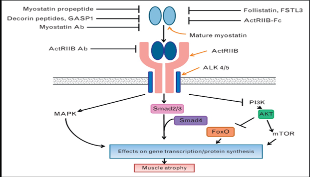
Fig. Summary of therapeutic invention points in the myostatin signaling pathway. Myostatin binds to its receptor complex ActRIIB/Alk 4 or 5 on skeletal muscle resulting in activation of the Smad 2/3, mitogen-activated protein kinase and inhibition of the PI3K intracellular signaling pathways that together result in gene transcriptional changes and effects on protein synthesis that ultimately give rise to muscle atrophy. Myostatin pathway inhibitors act extracellularly by either binding myostatin directly (Fstl3, Follistatin, myostatin antibody, GASP1, myostatin propeptide, decorin peptides, ActRIIB-Fc) or by binding its receptor complex (ActRIIB antibody) in order to block myostatin engaging its receptor complex and activating downstream signaling. Some of the inhibitors are naturally occurring (myostatin propeptide, Gasp1, follistatin, Fstl3) whereas others are engineered (myostatin antibody, ActRIIB antibody, ActRIIB-Fc). ---I represent inhibitory activities. → represent activating activities. Ab = antibody.
4. Genetic Modifiers and Myostatin
Myostatin has been identified as a genetic modifier of muscular dystrophies, including DMD. Variability in myostatin levels may influence the severity and progression of muscle degeneration in patients1. Understanding how genetic variations in the myostatin gene impact muscle function in DMD patients could inform personalized treatment approaches.
5. Future Research Directions
Ongoing research into myostatin’s role in muscle pathology will help clarify its mechanisms and interactions with other signaling pathways in DMD. Enhanced understanding of myostatin may lead to innovative therapies aimed at modulating its activity, potentially improving the quality of life and functional outcomes for patients with Duchenne muscular dystrophy.
“How Calcium Affects Muscle Health in Muscular Dystrophy”
Calcium plays a pivotal role in muscular functions, and its dysregulation is closely linked to the pathogenesis of muscular dystrophies, particularly Duchenne Muscular Dystrophy (DMD).
Calcium Handling in Muscle Function
Importance of Calcium in Muscle Contractions
Calcium is essential for muscle contractions as it facilitates the interaction between actin and myosin, the contractile proteins in muscle fibers. Calcium binds to troponin, which causes a conformational change in tropomyosin, exposing the binding sites on actin for myosin, resulting in muscle contraction.
Role in Cellular Processes
Calcium's role extends beyond muscle contraction. It is crucial for several other cellular processes, including nerve impulse transmission and blood clotting. Additionally, it helps in the release of neurotransmitters at synapses and the regulation of enzyme activities.
Calcium Dysregulation in Muscular Dystrophy
Duchenne Muscular Dystrophy (DMD)
Duchenne Muscular Dystrophy is characterized by a mutation in the DMD gene, leading to the absence of dystrophin, a protein crucial for maintaining muscle fiber integrity. The loss of dystrophin results in destabilized muscle cell membranes, making them susceptible to injury and disrupting calcium homeostasis.
Abnormal Calcium Handling
In DMD, the muscle cells exhibit dysregulated calcium handling due to several factors:
Abnormal Calcium Entry: The loss of dystrophin disrupts the sarcolemmal structure, facilitating abnormal calcium entry.
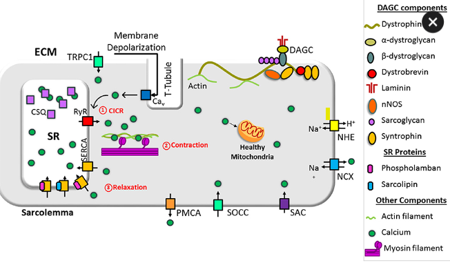
Sarcoplasmic Reticulum Dysfunction: There is increased release of calcium from the sarcoplasmic reticulum and impaired re-uptake due to dysfunctional SR Ca2+ ATPase (SERCA).
Mitochondrial Dysfunction: Dysregulation in calcium handling affects mitochondrial function, leading to elevated production of reactive oxygen species (ROS) and muscle damage.
Calcium as a Therapeutic Target
Current Research and Potential Therapies
Several studies suggest that targeting the calcium-handling mechanisms could be a promising therapeutic strategy for DMD. Researchers are exploring various approaches:
SERCA Activity ImprovementEnhancing SERCA function to normalize calcium re-uptake has shown potential in mitigating muscle damage in animal models.
Calcium Channel Modulators: Using inhibitors or modulators that regulate the abnormal influx of calcium into muscle cells is another area of active research.
Gene Therapy and Exon Skipping: These approaches aim to restore dystrophin or introduce functional substitutes like utrophin to stabilize muscle cell membranes and normalize calcium handling.
Conclusion
Calcium plays a critical role in muscle function and its dysregulation is Central to the pathology of Duchenne Muscular Dystrophy.Understanding the detailed mechanisms of calcium handling in muscle cells has opened up avenues for innovative therapeutic strategies targeting calcium homeostasis. Continuing research in this area holds promise for developing effective treatments to mitigate the progression of muscular dystrophies.
“Unraveling the Role of FOXO1 Signaling in Muscular Dystrophy: Pathways to Potential Therapies"
FOXO1 signaling plays a crucial role in muscle function by regulating muscle growth, metabolism, and atrophy. It influences the expression of various atrophy-related genes and is essential for maintaining muscle protein homeostasis, particularly under catabolic conditions. The inhibition or deletion of FOXO1 has been shown to protect against muscle loss during states of stress, such as fasting or denervation.
1. Regulation of Muscle Growth and Metabolism
FOXO1 is a key regulator of muscle growth, metabolism, cell proliferation, and differentiation1. This forkhead transcription factor mediates various metabolic pathways that are vital for maintaining muscle function, specifically through its influence on muscle energy homeostasis and the regulation of lipolytic and glycolytic fluxes.
2. Role in Muscle Atrophy
FoxO1 signaling contributes significantly to the onset of muscle atrophy. It has been observed that FOXO1, alongside other FOXO family members, is required to induce the expression of several atrophy-related genes during catabolic states. The absence of FOXO1 has been linked to a blunted induction of these genes, which are critical for muscle protein breakdown.
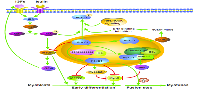
Fig.1. FoxO1 signaling pathway involved in skeletal muscle differentiation.
3. Impact of FoxO1 Deletion on Muscle Loss
FoxO1 inhibition plays a vital role in preventing muscle loss during periods of nutrient deprivation. Studies have shown that the deletion of FoxO1 in skeletal muscle protects against muscle loss and force decline in response to fasting. Furthermore, FOXO1 knockout mice showed complete protection from muscle mass reduction during fasting, highlighting its central function in muscle wasting
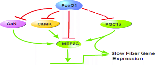
Fig.2 Mechanisms of FoxO1 in the regulation of slow skeletal muscle fibre gene expression. FoxO1 downregulates calcineurin (CaN), CaMK and MEF2C expression, leading to a decrease of MEF2C that can increase the transcriptional activation of slow fibre genes, to inhibit slow fibre genes expression
4. Interactions with Autophagy and Ubiquitin-Proteasome Systems
FOXO1 signaling is closely linked with autophagy and the ubiquitin-proteasome system, both crucial mechanisms for protein degradation in muscle cells. In conditions where nutrients are scarce, FOXO1 promotes the induction of autophagy and the activation of ubiquitin ligases, leading to the breakdown of muscle proteins. The disruption of FOXO1 activity impairs these degradation pathways, contributing to muscle protection.
5. Functional Implications of FOXO1 Deletion in Muscle
Research indicates that the absence of FOXO1 does not adversely affect muscle type determination or basal glucose homeostasis, suggesting its role is more critical during stress conditions. This regulatory pathway is essential for maintaining muscle mass and function, especially under catabolic stimuli like fasting or denervation, where FOXO1 expression contributes to the muscle atrophy program.
In summary, FOXO1 signaling is fundamental to muscle function, influencing growth, metabolism, and the process of muscle atrophy through complex interactions within the muscle tissue. Understanding these mechanisms, later on it can provide a therapeutic approach for muscle-wasting conditions.
"Interleukin-6: Friend or Foe in Muscular Dystrophy Management?"
Interleukin-6 (IL-6) is a pleiotropic cytokine involved in inflammation, immune response, and muscle metabolism. Its role in muscular dystrophy, particularly Duchenne Muscular Dystrophy (DMD), has been the subject of extensive research. This report delves into the mechanisms through which IL-6 impacts muscular dystrophy, its dual roles in muscle degeneration and regeneration, and the potential therapeutic strategies targeting IL-6.
IL-6 in Muscular Dystrophy
Chronic Elevation of IL-6 in DMD
IL-6 is chronically elevated in patients with Duchenne Muscular Dystrophy (DMD), a neuromuscular disorder characterized by the loss of the structural protein dystrophin. The absence of dystrophin leads to progressive muscle degeneration, chronic inflammation, and premature death. Elevated levels of IL-6 have been reported in both human DMD patients and dystrophin-deficient mdx mice, a commonly used animal model for human DMD.
Pro-Inflammatory Role and Muscle Degeneration
IL-6 is known to exacerbate muscle degeneration in DMD by sustaining the inflammatory response and promoting oxidative stress. Increased levels of IL-6 contribute to repeated cycles of muscle degeneration and regeneration, thereby amplifying tissue damage. Elevated IL-6 has been shown to worsen the dystrophic muscle phenotype by modulating inflammatory responses and playing a critical role in muscle maintenance and remodeling.
Functional and Morphological Effects
IL-6 exacerbates oxidative damage in dystrophic muscle, which can amplify degenerative processes. Increased serum levels of IL-6 are associated with muscle destruction and weakness in DMD patients. Studies have demonstrated that blocking IL-6 signaling can attenuate muscle damage and improve muscle function in dystrophin-deficient models, suggesting a potential therapeutic approach.
Mechanisms of IL-6 in Muscle Pathophysiology
Muscle Atrophy
IL-6 signaling is implicated in promoting muscle atrophy through several mechanisms. Recombinant IL-6 treatment in cultured C2C12 my tube cells increased the expression of atrophy-related genes, such as Atrogin1, and reduced the diameter of myotubes. The administration of IL-6 in mice induced the loss of muscle mass, inhibited the mammalian target of rapamycin (mTOR) pathway, and reduced phosphorylation of eukaryotic translation initiation factor 4E-binding protein 1 (4E-BP1), thus diminishing protein synthesis and activating muscle atrophy mechanisms.
Muscle Regeneration and Myogenesis
Despite its atrophic effects, IL-6 also promotes muscle regeneration and myogenesis. IL-6 signaling has been associated with hypertrophic muscle growth and the regulation of muscle cell proliferation11. This dual role complicates the therapeutic targeting of IL-6, as it is involved both in muscle degeneration and in the regenerative processes that are critical for muscle repair.
IL-6 as a Therapeutic Target
Due to its significant role in muscle pathology, IL-6 has emerged as a potential therapeutic target for muscular dystrophy. Anti-IL-6 receptor antibodies have been shown to improve muscle destruction and weakness in animal models of DMD. These findings suggest that inhibiting IL-6 signaling could mitigate inflammation and muscle degeneration, thus improving muscle function and slowing disease progression.
Clinical Trials and Research
Recent studies and clinical trials have focused on the efficacy of IL-6 blockade in mitigating muscular dystrophy symptoms. A study conducted in 2023 indicated elevated IL-6 levels in the serum and cerebrospinal fluid of DMD patients, underscoring the cytokine's role in the disease's pathology. Such research highlights the therapeutic potential of modulating IL-6 activity in DMD treatment strategies.
Conclusion
Interleukin-6 is an important cytokine in the context of muscular dystrophy, particularly Duchenne Muscular Dystrophy. Its elevated levels contribute to chronic inflammation, muscle degeneration, and oxidative stress, while also playing a role in muscle regeneration. Therapeutic strategies targeting IL-6 signaling have shown promise in preclinical studies, offering potential avenues for mitigating muscle damage and improving patient outcomes. Further research is essential to fully understand the complex role of IL-6 and to develop effective interventions for muscular dystrophy.
"The Role of Matrix Metalloproteinase in Muscle Remodelling and Muscular Dystrophy"
Matrix metalloproteinase (MMPs) are the enzymes that play very significant roles in muscle remodeling and the pathogenesis of muscular dystrophy.Their activities are essential for maintaining extracellular matrix (ECM) integrity, facilitating muscle regeneration, and influencing inflammatory processes. Understanding the functions of MMPs and their regulation, opens potential ways for therapeutic strategies aimed to treat muscular dystrophies.
IL-6 in Muscular Dystrophy
1. Role in Muscle Remodelling
MMPs are regulatory proteases involved in synthesizing, degrading, and remodeling extracellular matrix (ECM) components. By modulating ECM reconstruction, cellular migration, and differentiation, MMPs preserve myofiber integrity and homeostasis, which is critical for muscle function. They contribute to the degradation of collagen, proteoglycans, and other ECM components, enabling muscle regeneration and repair. MMPs also facilitate the recruitment of inflammatory cells to sites of tissue damage, further assisting in muscle remodeling and recovery.
2. Involvement in Muscular Dystrophy
In muscular dystrophy, particularly Duchenne muscular dystrophy (DMD), MMPs—especially MMP-2 and MMP-9—are significantly involved in the disease's pathogenesis. MMP-9 has been shown to play a role in the inflammatory process during muscle degeneration. MMP-2 is associated with ECM remodeling during muscle regeneration. The dysregulation of MMP activities contributes to muscle degeneration and impaired regeneration in dystrophic conditions.
3. Impact on Muscle Repair and Regeneration
MMPs are essential for muscle regeneration and repair by degrading ECM components and facilitating satellite cell migration and differentiation. Specifically, MMP-1 aids in muscle healing by digesting fibrous scar tissue that can obstruct regeneration. Furthermore, regular exercise has been shown to increase MMP expression, which supports muscle repair and regeneration processes. The activation of MMPs through signaling pathways, such as TGF-β, is crucial for muscle regeneration.
4. Relationship with Inflammation
The involvement of MMPs in inflammation is notable in the context of muscular dystrophies. MMP upregulation is associated with inflammatory processes, and their elevation in muscular dystrophies results partly from inflammation. The role of MMPs in ECM breakdown and remodeling highlights their contribution to both degeneration and inflammation, with potential consequences for muscle recovery and adaptation.
5. Therapeutic Approaches
Given their significant involvement in muscle pathology, MMPs present potential therapeutic targets for muscular dystrophies. Inhibition of MMP-9 has been shown to improve skeletal muscle regeneration in dystrophic models. MMP inhibitors, such as batimastat, have demonstrated promise in preclinical studies for eradication of disease progression in DMD. Exploring the therapeutic potential of MMP modulation could lead to new interventions aimed at enhancing muscle regeneration and reducing fibrosis in muscular dystrophy.
"Targeting PTEN: A New Hope for Duchenne Muscular Dystrophy Therapy?"
PTEN (Phosphatase and tensin homolog) serves as a key regulatory protein in muscle growth and pathology, particularly in Duchenne muscular dystrophy (DMD). Its inhibition has gained attention as a promising therapeutic strategy to mitigate muscle degeneration and enhance muscle function in DMD models. Research shows that targeting PTEN can lead to significant improvements in muscle integrity and strength, presenting a new pathway for treatment.
1. Role of PTEN in Muscle Regulation
PTEN acts as a tumor suppressor and a negative regulator of muscle growth. In the context of DMD, studies have shown that the levels of PTEN increase considerably, which correlates with muscle degeneration and impaired muscle repair mechanisms. The inhibition of PTEN, whether through genetic knockout or pharmacological means, has demonstrated improvements in muscle function and pathology in mouse models of DMD.
2. Therapeutic Approaches Targeting PTEN
Recent research has explored various methods for inhibiting PTEN in DMD. Genetic and pharmacological approaches targeting PTEN have been shown to reduce skeletal muscle injury and inflammation while improving membrane repair. These findings highlight the therapeutic potential of PTEN inhibition as a strategy to enhance muscle recovery and limit degeneration associated with DMD.
3. Evidence from DMD Models
In DMD models, lowering PTEN levels has yielded promising results, allowing for larger and stronger muscle growth. In healthy muscle, PTEN levels are low; however, elevated levels in DMD are linked to muscular injury and dysfunction. Research is underway to develop safe and efficient pharmacological methods to deliver PTEN inhibitors specifically to skeletal muscle, aiming to boost muscle regeneration and strength.
4. Broader Implications of PTEN Inhibition
The findings surrounding PTEN's role in DMD extend to broader implications for understanding muscle degeneration in various conditions. New therapeutic strategies based on PTEN modulation offer insights into the mechanistic pathways that govern muscle integrity and regeneration. As research progresses, these strategies could lead to novel clinical treatments aimed at improving quality of life for individuals with DMD and potentially other muscular disorders
5. Future Directions in Research
Future research efforts are focused on mechanistically characterizing how direct inactivation of PTEN in muscle cells benefits DMD patients. Studies aim to elucidate molecular pathways involved in muscle hypertrophy and recovery in DMD by manipulating PTEN levels. By establishing robust preclinical models, researchers strive to validate PTEN as a target for therapeutic intervention in muscular dystrophies.
"How PINK1 is Revolutionizing Muscular Dystrophy Research and Treatment"
PINK1 (PTEN-induced kinase 1) plays a significant role in the pathophysiology of muscular dystrophy, particularly through its involvement in the mitophagy process and maintaining mitochondrial health. Dysregulation of the PINK1/Parkin pathway is associated with defective mitophagy in muscular dystrophy, leading to compromised muscle cell integrity and function.
1. Role of PINK1 in Mitophagy
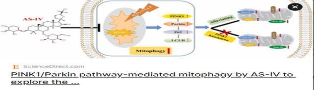
PINK1 is essential for the regulation of mitophagy, which is a mitochondrial quality control process that removes damaged mitochondria to protect cells from degeneration. The PINK1/Parkin pathway is specifically implicated in the degradation of dysfunctional mitochondria, an important mechanism in muscles that require high energy levels for contraction. In muscular dystrophies, defects in this pathway have been observed, resulting in an accumulation of damaged mitochondria and subsequent muscle fiber damage.
2. Impaired PINK1/Parkin Pathway in Muscular Dystrophy
Research indicates that mitochondrial pathology in dystrophic muscles is closely linked to malfunctions in the PINK1/Parkin-mediated mitophagy. Studies have shown that faulty mitophagy in dystrophic hearts is due to defects in this pathway, which underscores the critical nature of PINK1 in maintaining muscle health. The inability to effectively degrade damaged mitochondria contributes to muscle degeneration seen in conditions like Duchenne muscular dystrophy (DMD).
3.Therapeutic Implications
Given PINK1’s role in mitophagy and muscle integrity, targeting the PINK1/Parkin pathway presents a possible therapeutic strategy for muscular dystrophies. By enhancing the functionality of this pathway, it may be possible to improve mitochondrial quality control, thus preserving muscle fibers and improving overall muscle function. Research into PINK1’s pathways may lead to novel therapeutic interventions aimed at counteracting muscle degeneration associated with dystrophies.
4. Evidence from Experimental Models
Studies utilizing animal models of muscular dystrophy have indicated that PINK1 activity is crucial for muscle maintenance. The PINK1/Parkin pathway is often explored in experiments to investigate its implication in muscle repair and regeneration, emphasizing the necessity of functional mitophagy in preventing muscle degradation. Such findings highlight that therapeutic strategies focusing on enhancing PINK1 signaling could directly impact muscle pathology.
5. Future Directions in Research
Further research is warranted to elucidate the precise mechanisms by which PINK1 and its associated pathways contribute to muscular dystrophy. Investigating the potential for small molecule enhancers of PINK1 function, as well as gene therapy approaches, could further advance the understanding and treatment of muscular dystrophies. A multi-faceted approach targeting PINK1’s role in muscle biology may yield significant improvements in managing dystrophic diseases.
By consolidating the understanding of PINK1's functions and improving targeted therapies, it may be possible to mitigate some of the severe consequences associated with muscular dystrophies.
"Harnessing mTORC1 in the Fight Against Muscular Dystrophy"
Overview of mTORC1 Function
mTORC1 is critically important in the regulation of skeletal muscle mass through its regulation of protein synthesis and degradation pathways. mTORC1 is activated by various stimuli such as mechanical load, amino acids, and growth factors. It regulates protein synthesis by activating S6K1 and inhibiting 4EBP1, promoting muscle growth.
Dysregulation of mTORC1 in Muscular Dystrophies
Duchenne Muscular Dystrophy (DMD)
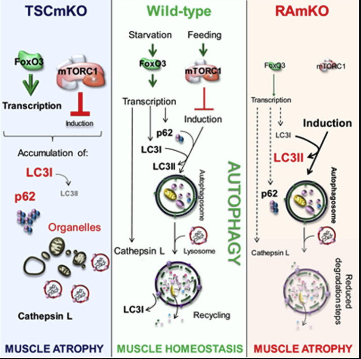
Enhanced Akt-mTORC1 signaling has been shown to impair autophagy and contribute to muscular and cardiac damage in Duchenne Muscular Dystrophy (DMD). Moreover, activation of mTORC1, inhibition of autophagy, and the accompanying muscle weakness on the loss of dystrophin have been observed in mdx mice, a model for DMD.
Dystroglycanopathy
mTORC1 signaling also correlates with levels of pathological markers in dystroglycanopathy muscle, emphasizing its role in disease pathology. Dysregulation of mTORC1 in these conditions leads to impaired autophagy and subsequent muscle degeneration.
mTORC1 and Myopathy
Sustained activation of mTORC1 through TSC1 deficiency leads to a late-onset myopathy related to impaired autophagy. In muscle-specific mTOR knockout mice, severe myopathy is observed, indicating the critical role of mTOR in muscle function.
Therapeutic Strategies Targeting mTORC1
mTORC1 Inhibitors
Rapamycin: Inhibition of mTORC1 with rapamycin and related compounds has shown promise in treating muscular dystrophies. Rapamycin treatment improved muscle relaxation and increased muscle force in HSALR mice, a model for dominant muscular dystrophy. Additionally, rapamycin has been demonstrated to reduce muscle necrosis and T cell infiltration in mdx mice.
Autophagy Modulation
Given the critical role of mTORC1 in regulating autophagy, therapeutic approaches that modulate autophagy may benefit muscular dystrophy patients. Enhancing autophagy through mTORC1 inhibitors like rapamycin could potentially mitigate muscle degeneration by promoting the clearance of damaged proteins and organelles.
Conclusion
The mTORC1 signaling pathway is a critical regulator of muscle mass and function, with significant implications in the pathogenesis and treatment of muscular dystrophies. Dysregulation of this pathway contributes to disease progression, and targeting mTORC1 signaling represents a promising therapeutic strategy. Continued research is essential to fully understand the complexities of mTORC1 signaling in muscular dystrophies and to develop effective treatments.
"Dystroglycanopathy: Exploring the Genetic and Molecular Landscape of Muscular Dystrophy"
Dystroglycanopathy represents a group of inherited muscular dystrophies characterized by hypoglycosylation of the extracellular protein α-dystroglycan. These disorders significantly affect muscle integrity, often leading to severe muscle weakness and other associated conditions impacting the central nervous system and eyes. Due to the genetic and phenotypic variability of dystroglycanopathy, accurate diagnosis remains a challenge.
1. Overview of Dystroglycanopathy
Dystroglycanopathy is a collective term for muscular dystrophies caused by abnormal glycosylation of dystroglycan, an integral component of the dystrophin-glycoprotein complex2. These disorders lead to a spectrum of muscular dystrophies and can present with additional neurological and ocular defects. Alpha-dystroglycan is essential for maintaining the structural integrity of muscle cells by linking the intracellular cytoskeleton to the extracellular matrix.
2. Genetic Basis and Variants
The genetic basis of dystroglycanopathy is quite complex, involving mutations in several genes that encode proteins responsible for the glycosylation of α-dystroglycan. Currently18 genes have been associated with dystroglycanopathies, with variations in these genes leading to diverse clinical presentations and severity of symptoms. Understanding the specific mutations is crucial for accurate diagnosis and potential treatment strategies.
3. Clinical Features
Patients with dystroglycanopathies generally exhibit proximal muscle weakness, elevated serum creatine kinase levels, and various forms of limb-girdle muscle weakness. The age of onset can vary significantly, and the clinical manifestations may range from severe congenital forms, such as Walker-Warburg syndrome, which include brain and eye anomalies, to milder forms that emerge in adulthood without central nervous system involvement. This variability complicates both diagnosis and treatment approaches.
4. Diagnostic Techniques
Accurate diagnosis of dystroglycanopathy has historically been challenging due to its clinical and genetic heterogeneity. Next-generation sequencing (NGS) techniques, including whole-exome sequencing (WES), are increasingly employed to identify pathogenic variants in dystroglycanopathy-associated genes. The ability to rapidly sequence the exome allows for a clearer understanding of the underlying genetic mechanisms and aids in guiding therapeutic interventions.
5. Current Treatment Strategies
As of now, there are no specific therapies that can halt the progression of dystroglycanopathies. Current approaches primarily focus on managing symptoms and improving quality of life through physical therapy and supportive care. Research continues to explore innovative therapeutic strategies, including gene therapy and pharmacological interventions that could target the underlying genetic and biochemical abnormalities associated with these conditions.
6. Research and Future Directions
Ongoing research is vital to fully characterizing the molecular mechanisms of dystroglycanopathies and identifying new therapeutic targets. Investigations into the genetic diversity and functional consequences of mutations will be fundamental in developing effective treatment plans tailored to individual patients. Ultimately, creating a comprehensive international registry for dystroglycanopathy could enhance data collection and promote collaborative research efforts.
In summary, dystroglycanopathies represent a complex and diverse group of disorders requiring thorough genetic analysis for effective diagnosis and management, with ongoing research focused on improving understanding and treatment options
"The Immune Connection: How CD8+ T Cells Influence Muscular Dystrophy Progression"
CD8+ T cells play a important role in the pathogenesis of muscular dystrophies, particularly in Duchenne Muscular Dystrophy (DMD) and other inflammatory myopathies. Their activation and subsequent immune responses can contribute to muscle inflammation and damage, ultimately affecting muscle regeneration. Understanding the dynamics of CD8+ T cells in these conditions provides insights into potential therapeutic approaches aimed at modulating the immune response.
1. Role of CD8+ T Cells in Muscle Damage
CD8+ Tcells, also known as cytotoxic T cells, are implicated in the muscle damage
observed in muscular dystrophies like Duchenne Muscular Dystrophy (DMD). They are activated in response to muscle injury and participate in the immune response by targeting and eliminating damaged or diseased muscle fibers. This activation can lead to inflammation, contributing to the deleterious cycle of muscle degeneration and impaired regeneration.
2. Immune Activation in Duchenne Muscular Dystrophy
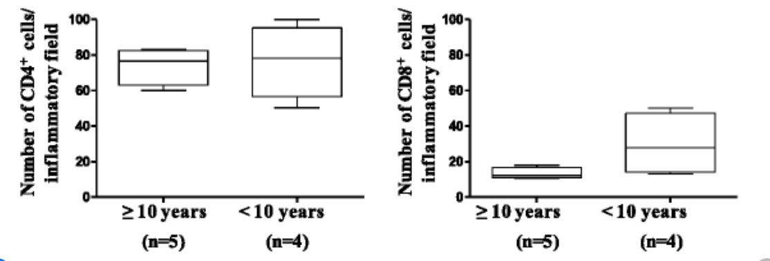
In boys with Duchenne Muscular Dystrophy, there is evidence of complex immune
activation involving increased numbers of circulating CD8+ T cells. These cells show enhanced binding of adenosine deaminase, which
correlates with higher muscular strength scores, signifying a relationship
between immune cell activation and muscle functionality. This suggests that while
CD8+ T cells may contribute to muscle damage, their role may also be essential in the muscle repair process under specific conditions.
3. Consequences of CD8+ T Cell Activity
The cytotoxic activity of CD8+ T cells can lead to increased myocyte apoptosis and
subsequent muscle fiber degeneration. The interplay between these immune cells and muscle progenitor cells is crucial, as excessive activation without appropriate regulation can exacerbate the disease progression1. Therefore,Understanding the balance of CD8+ T cell activity is vital for developing targeted therapies in muscular dystrophy.
4. Implications for Therapeutic Strategies
Targeting CD8+ T cell responses holds potential for therapeutic interventions in muscular
dystrophies. Modulating the immune response, either by inhibiting excessive
cytotoxic activity or enhancing regulatory pathways, may mitigate muscle damage
and improve regeneration. This approach could lead to improved treatment outcomes, addressing both the inflammatory and degenerative aspects of muscular dystrophy.
5. Future Research Directions
Ongoing research is focused on elucidating the specific mechanisms through which CD8+ T cells influence muscle pathophysiology. Investigating the potential of
interventions that could balance CD8+ T cell activity without compromising
immune protection may present novel avenues for treatment. Such strategies could offer
hope in enhancing muscle function and quality of life for individuals affected
by muscular dystrophies.