Hathiyar,Bodh Gaya Mon-Sat 09am-7pm, Sun Closed
Call Now
+91-748887920
exRNARare
Mail Now
info@exrnarare.com
Sanchi Singh

Research Assistant, exRNA Therapeutics


"Kartagener Syndrome and Respiratory Tract Dysfunction: A Comprehensive Overview”

Kartagener syndrome is a rare hereditary disorder that is inherited in autosomal recessive consisting a ‘clinical triad’ with chronic sinusitis, bronchiectasis and situs inversus syndrome. The prevalence of primary ciliary dyskinesia varies from 1/20 000 to 1/60 000 live births. Kartagener’s syndrome results due to primary ciliary dyskinesia (PCD) leading to decreased mucosal scavenging capability and increased infection susceptibility in the respiratory tract that leads to symptoms like progressive symptoms like productive cough, wheezing, and shortness of breath.


Occasionally Kartagener’s syndrome can be associated with reversible airflow obstruction which is managed with the prescription of anti-asthmatics like steroids and beta-adrenergic agonists. People with this condition have a high risk of getting recurrent bacterial and fungal infections


ORIGIN

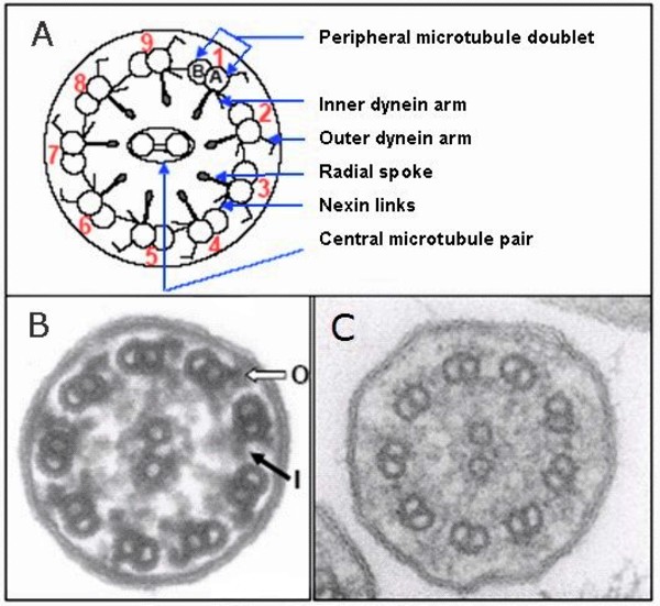
As it is a genetic disease, the cause of Kartagener syndrome is a mutation in different genes associated with the alteration of the cilia. Although DNAH 11 a gene encode a protein that is a part of the outer dynein arm of cilia and is required for cilia motility and CCDC40, responsible for assembly of internal dynein arm.Mutations in these genes cause the loss of the dynein arms of the cilia and thus prevent their movement.

Italian Trulli


A) schematic diagram of a cilium; B) electron microscopic image of a normal cilium with "O" representing the outer dynein arm and "I" representing the inner dynein arm; C) electron microscopic image of a cilium of an individual with primary ciliary dyskinesia showing absence of dynein arms

IMPACT ON RESPIRATORY TRACT

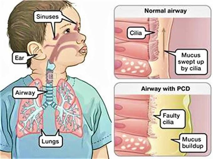
In the respiratory tract, cilia move back and forth in a coordinated way to move mucus towards the throat. This movement of mucus helps to eliminate fluid, bacteria, and particles from the lungs. Most babies with primary ciliary dyskinesia experience breathing problems at birth, which suggests that cilia play an important role in clearing fetal fluid from the lungs. Beginning in early childhood, affected individuals develop frequent respiratory tract infections. Without properly functioning cilia in the airway, bacteria remain in the respiratory tract and cause infection. People with primary ciliary dyskinesia also have year-round nasal congestion and a chronic cough. Chronic respiratory tract infections can result in a condition called bronchiectasis, which damages the passages, called bronchi, leading from the windpipe to the lungs and can cause life-threatening breathing problems.

Italian Trulli


Fig showing Normal airway cilia in compare with Airway with Primary Ciliary Dyskinesia

REFERENCES 1 : Click Me
REFERENCES 2 : Click Me

Sanchi Singh

Research Assistant, exRNA Therapeutics


Understanding Activated PI3K Delta Syndrome (APDS): A Rare Primary Immunodeficiency

Activated PI3K delta syndrome (APDS) is a rare primary immunodeficiency (PI) first identified in 2013. This condition is caused by genetic variants in one of two genes: PIK3CD or PIK3R1. These genes play a crucial role in the development and function of immune cells, making them essential for a healthy immune system.


Causes of APDS

APDS arises from mutations in either the PIK3CD or PIK3R1 genes, leading to an overactive enzyme called phosphoinositide 3-kinase delta (PI3K delta). This enzyme is vital for the normal development and proliferation of B-cells and T-cells key components of the immune system. However, in APDS, the overactivity of PI3K delta disrupts the normal functioning of these cells, compromising the body’s ability to fight infections.

There are two types of APDS:

  • APDS1: Caused by mutations in the PIK3CD gene.
  • APDS2:: Caused by mutations in the PIK3R1 gene.
Symptoms of APDS

APDS can present a wide range of symptoms, making diagnosis challenging. Common early symptoms include frequent and severe infections of the ears, sinuses, and respiratory tract. Other symptoms may include:

  • Chronic cough
  • Enlarged tonsils, lymph nodes, and spleen
  • Nodules in the airways and digestive tract
  • Gastrointestinal issues
  • Autoimmune and autoinflammatory disorders
  • Herpes infections
  • Low blood cell counts
  • Lymphoma
  • Developmental delays
Diagnosis of APDS

The only definitive way to diagnose APDS is through genetic testing. Given that PIs are inherited, it’s crucial for family members of a confirmed patient to undergo genetic testing as well. Even if they don’t show symptoms, they may carry the genetic variant and could pass it on to their children.


Treatment Options for APDS

Treatment for APDS is tailored to the individual’s symptoms. Infections are typically managed with antibiotics, antifungals, or antiviral medications. For those prone to recurrent infections, ongoing preventive (prophylactic) treatments may be necessary. Patients with poor immunoglobulin (antibody) production may benefit from immunoglobulin replacement therapy. Additionally, drugs like steroids, sirolimus (an mTOR inhibitor), or the monoclonal antibody rituximab can be used to modulate the immune response, reduce spleen or lymph node size, and improve blood counts. Emerging therapies specifically targeting the overactive PI3K delta enzyme known as PI3K delta inhibitors have shown promising results, particularly in cases with excessive white blood cell production (lymphoproliferation). For some, hematopoietic stem cell transplantation (HSCT), also known as bone marrow transplantation (BMT), offers a potential cure for APDS.


Conclusion

While APDS is a complex and rare condition, understanding its causes, symptoms, and treatment options can lead to better management and outcomes for patients. If you or a loved one is affected by APDS, staying informed and working closely with healthcare providers is key to navigating this challenging condition.


Shivam Yadav

Research Assistant (exRNA therapeutics)


Amyotrophic Lateral Sclerosis (ALS): Pioneering Antisense Oligonucleotide Therapy for a Rare Neurodegenerative Disease

Amyotrophic Lateral Sclerosis (ALS), also known as Lou Gehrig's disease, is a devastating neurodegenerative disorder that progressively robs individuals of their ability to move, speak, and ultimately breathe. Affecting an estimated 2 in 100,000 people globally each year, ALS is classified as a rare disease, though its impact on patients and families is profound and widespread. Despite extensive research, effective treatment options have been limited, driving an urgent need for innovative therapies.


At the forefront of this battle is the emerging field of antisense oligonucleotide (ASO) therapy, offering new hope in the quest to treat this debilitating condition. Our organization is deeply committed to advancing ASO-based treatments, harnessing the power of genetic medicine to target the root causes of ALS.


Understanding ALS: A Complex Neurodegenerative Disorder

ALS is characterized by the progressive degeneration of motor neurons in the brain and spinal cord. These neurons are responsible for transmitting signals from the brain to the muscles, enabling voluntary movements. As these neurons deteriorate, patients experience muscle weakness, spasticity, and eventually paralysis. While cognitive function is typically preserved, the loss of motor function is relentless, leading to death within 3 to 5 years of diagnosis for most patients.


The causes of ALS are multifactorial, with a combination of genetic and environmental factors at play. About 10% of cases are familial, linked to mutations in specific genes such as SOD1, C9orf72, and FUS. The remaining 90% are sporadic, with no clear hereditary component. Despite this variability, the underlying pathology involves the accumulation of toxic proteins, oxidative stress, mitochondrial dysfunction, and inflammation, all contributing to motor neuron death.

The Promise of Antisense Oligonucleotide Therapy in ALS

Antisense oligonucleotides (ASOs) represent a promising therapeutic approach for ALS by directly targeting the genetic mutations and molecular pathways implicated in the disease. ASOs are short, synthetic strands of nucleotides designed to bind to specific RNA sequences, modulating gene expression or correcting abnormal RNA splicing. This precision allows ASOs to silence harmful genes, promote the degradation of toxic proteins, or restore normal protein function.


In the context of ALS, ASOs have shown particular promise in targeting known genetic mutations. For instance, ASOs targeting the SOD1 gene, which accounts for about 2% of all ALS cases, have demonstrated the ability to reduce the production of toxic SOD1 protein aggregates in preclinical models. Clinical trials with SOD1-targeted ASOs, such as tofersen, have shown encouraging results, slowing disease progression in patients with SOD1 mutations


Another promising target is the C9orf72 gene, which harbors an abnormal hexanucleotide repeat expansion responsible for up to 40% of familial ALS cases. ASOs designed to reduce the toxic RNA foci and dipeptide repeat proteins produced by this mutation are currently in development, with early-phase trials indicating potential benefits.

Overcoming Challenges in ASO-Based Therapy

While ASO therapy holds significant promise, several challenges remain. Delivering ASOs to the central nervous system (CNS) is complex, requiring intrathecal administration to bypass the blood-brain barrier. Additionally, the long-term safety and efficacy of ASOs need further investigation, particularly in light of potential immune responses and off-target effects.


To address these challenges, our organization is dedicated to refining ASO design, optimizing delivery methods, and conducting rigorous clinical trials. By focusing on precision medicine, we aim to develop ASOs that not only target the genetic underpinnings of ALS but also offer personalized treatment options tailored to individual patient profiles.


The Future of ALS Treatment: A New Era of Hope

The development of ASO-based therapies marks a new era in the treatment of ALS, offering the possibility of slowing or even halting the progression of this relentless disease. As research advances, we are hopeful that ASOs will become a cornerstone of ALS treatment, transforming the lives of patients and their families.


Our organization remains at the forefront of this exciting field, committed to driving innovation and collaboration in the pursuit of effective treatments for ALS and other rare neurodegenerative diseases. By continuing to push the boundaries of genetic medicine, we aim to bring new hope to those affected by ALS, paving the way for a future where this devastating disease is no longer a death sentence but a manageable condition.


REFERENCES 1 : Click Me
REFERENCES 2 : Click Me
REFERENCES 3 : Click Me

SANCHI SINGH

Research Assistant (exRNA therapeutics)


When Lungs Collapse: A Deep Dive into Spontaneous Pneumothorax and Its Management


Spontaneous pneumothorax refers to the abnormal collection of gas in the pleural space between the lungs and the chest wall. Spontaneous pneumothorax occurs without an obvious etiology such as trauma or iatrogenic causes. Spontaneous pneumothorax can be classified as either primary or secondary. Primary spontaneous pneumothorax (PSP) occurs when the patient does not have a history of the underlying pulmonary disease, whereas secondary spontaneous pneumothorax (SSP) is associated with a history of an underlying pulmonary disease.


Etiology

Primary Spontaneous pneumothorax is associated with mutation in the FLCN gene.


  • Mutations in the FLCN gene can cause primary spontaneous pneumothorax, although these mutations appear to be a very rare cause of this condition.
  • The FLCN gene provides instructions for making a protein called folliculin. In the lungs, folliculin is found in the connective tissue cells that allow the lungs to contract and expand when breathing. Folliculin is also produced in cells that line the small air sacs (alveoli).
  • Familial PSP (10-20% of cases)
  • Genetic syndromes:- Marfan syndrome (FBN1 mutation) - Ehlers-Danlos syndrome (COL3A1 mutation) - Cystic fibrosis (CFTR mutation) - Neurofibromatosis type 1 (NF1 mutation)
Secondary spontaneous pneumothorax is associated with underlying pulmonary diseases, but not limited to the following:
  • Chronic obstructive pulmonary disease
  • Asthma
  • Cystic fibrosis
  • Pneumonia
  • Pulmonary abscess
  • Tuberculosis
  • Malignancy
  • Interstitial lung disease (e.g., idiopathic pulmonary fibrosis, sarcoidosis, lymphangioleiomyomatosis)
  • Connective tissue disease (e.g., Marfan syndrome, Ehlers-Danlos syndrome, rheumatoid arthritis)
  • Pulmonary infarct
  • Foreign body aspiration
  • Catamenial (i.e., associated with menses secondary to thoracic endometriosis)
  • Birt-Hogg-Dube syndrome

Evaluation

The diagnosis of spontaneous pneumothorax is often suggested by the patient’s history and physical exam findings, which can be confirmed b

  • Imaging
  • CT (Chest Tomography)
  • Chest Radiography
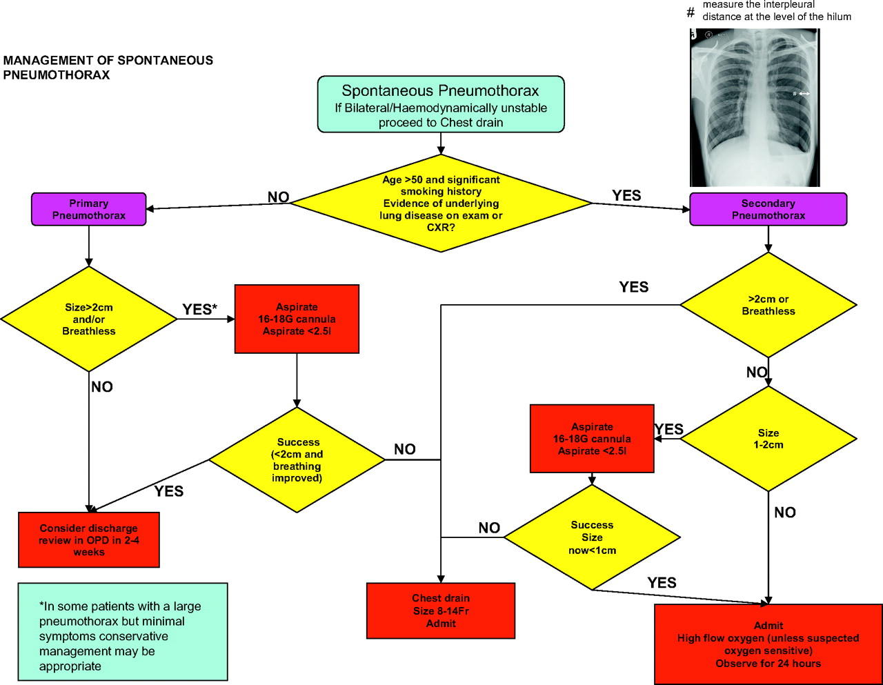
Treatment / Management
  • The main goal for the treatment of spontaneous pneumothorax is to evacuate the gas from the pleural space and the prevention of recurrences
  • Nevertheless, it is appropriate to initiate 100% oxygen via a non-rebreather mask and continuous cardiopulmonary monitoring for patients with spontaneous pneumothorax. Oxygen increases the rate of absorption of the gas from the pleural space up to four-fold compared to the absorption of 1% to 2% of the volume per day without oxygen. Clinically unstable patients with severe symptoms or symptoms suggestive of tension pneumothorax can be treated with emergent needle decompression as a bridge to tube thoracostomy placement
  • Patients with recurrent primary spontaneous pneumothorax should be admitted with thoracostomy tube placement as a bridge to VATS. In patients that are unwilling to undergo VATS, are poor surgical candidates, or are being managed in an institution where VATS is not readily available, chemical pleurodesis can be performed with the introduction of irritants such as tetracyclines (i.e., doxycycline, minocycline) or talc via the thoracostomy tube. The inflammatory processes associated with chemical pleurodesis lead to the formation of pleural adhesions that effectively obliterate the pleural space.
  • Patients with recurrent secondary spontaneous pneumothorax re recommended with supplemental oxygen and repeat chest radiograph ( in small pneumothorax), placement of a pleural catheter or thoracostomy tube ( in lage pneumothorax)the patient is symptomatic, or the secondary spontaneous pneumothorax is bilateral.
  • Observation alone is not recommended as there is an increased risk for mortality in secondary spontaneous pneumothorax. Referral to a thoracic specialist is recommended, but not until the patient is stabilized with a chest drain.

Italian Trulli

Italian Trulli


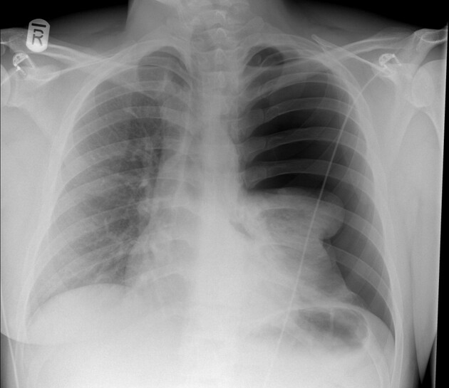
Large tension pneumothorax on the left side is noted with near complete collapse of the left lung and mild mediastinal shift towards right side.


REFERENCES 1 : Click Me
REFERENCES 2 : Click Me
REFERENCES 3 : Click Me
REFERENCES 4 : Click Me