Research Assistant, exRNA Therapeutics
Arterial Tortuosity Syndrome (ATS) is a rare but profoundly serious genetic disorder that disrupts the integrity of the body’s arterial system. Characterized by the abnormal twisting, elongation, and weakening of the arteries, ATS poses significant risks to affected individuals, often leading to life-threatening complications such as aneurysms, stenosis, and dissections. This disorder demands careful attention from both the medical community and families affected by it, as its implications can be severe and far-reaching.
ATS is inherited in an autosomal recessive pattern, requiring two copies of a mutated gene—one from each parent—for the disorder to manifest. The primary gene implicated in ATS is SLC2A10, which encodes the GLUT10 protein. GLUT10 plays a role in cellular processes, but when this gene is mutated, it disrupts the normal development of connective tissue, particularly in the arteries. This leads to weakened arterial walls that are prone to the twisting and elongation characteristic of ATS. The genetic basis of ATS not only defines the condition but also complicates its management. Because ATS is rare, with fewer than a few hundred cases reported globally, understanding the full spectrum of its genetic mutations and their consequences is still an ongoing process. This makes timely diagnosis and tailored treatment plans critical, yet challenging.
The hallmark of ATS is arterial tortuosity, which can lead to a range of serious complications. These include:
Aneurysms: The abnormal bulging of arterial walls due to their weakened structure is a frequent and potentially fatal complication. If an aneurysm ruptures, it can cause severe internal bleeding, requiring emergency intervention.
Stenosis: Narrowing of the arteries can restrict blood flow to critical organs and tissues, leading to ischemia, pain, and even organ failure.
Arterial Dissections: Tearing of the artery’s inner layer can create a life-threatening situation, where blood is diverted into a false channel within the artery wall, potentially leading to stroke, heart attack, or organ damage.
In addition to these vascular issues, ATS often presents with systemic features such as hyperextensible skin, joint hypermobility, and distinctive facial characteristics, further complicating the lives of those affected. These symptoms can significantly impair quality of life and require a multidisciplinary approach to management.
The diagnosis of ATS is a critical step in managing the condition, but it is often delayed due to its rarity and the variability of its symptoms. Patients may present with signs of vascular complications that are mistaken for other, more common conditions. However, the identification of ATS is crucial because early diagnosis allows for the implementation of monitoring and preventive measures that can mitigate the risk of life-threatening complications.
Genetic Testing: Identifying mutations in the SLC2A10 gene confirms the diagnosis and can inform family planning decisions. Imaging Studies: Advanced imaging techniques, including MRI, CT angiography, and echocardiography, are essential for visualizing the extent of arterial tortuosity and detecting aneurysms, stenosis, or dissections early.
Managing ATS is an ongoing challenge due to the lack of curative treatments. Current medical care focuses on mitigating risks and addressing complications as they arise.
Regular Surveillance: Frequent imaging studies are necessary to monitor the progression of arterial tortuosity and identify any new or enlarging aneurysms before they become critical. Blood Pressure Management: Strict control of blood pressure is essential to reduce the stress on already weakened arteries, lowering the risk of aneurysm rupture and dissection.
Complex Surgeries: When surgical intervention is required, such as for aneurysm repair, the procedure is often complex and risky due to the fragile and tortuous nature of the arteries in ATS patients. Surgeons must balance the need to intervene with the risks of surgery itself, making these decisions particularly challenging.
Endovascular Approaches: In some cases, less invasive endovascular techniques may be used to manage aneurysms or stenoses, but these also carry significant risks due to the fragility of the arteries.
Lack of Disease-Modifying Therapies: One of the most serious issues in ATS management is the absence of treatments that can alter the course of the disease. Current approaches are primarily supportive and preventive, rather than curative.
The rarity of ATS contributes to the challenges in developing effective treatments. With limited patient populations, research funding, and clinical trials, progress has been slow. However, the seriousness of the condition underscores the urgent need for continued research into the molecular mechanisms underlying ATS, with the goal of developing targeted therapies that could prevent or even reverse the damage to arterial walls.
Gene Therapy: Advances in gene therapy offer hope for addressing the root cause of ATS by correcting the genetic mutations responsible for the disorder. Molecular Research: Understanding how SLC2A10 mutations lead to vascular abnormalities could open new avenues for therapeutic intervention, potentially leading to treatments that strengthen arterial walls and prevent tortuosity.
For those living with ATS, the burden of the disease extends beyond medical complications. The psychological and emotional impact of managing a serious, lifelong condition cannot be overstated. Access to comprehensive care teams, including cardiologists, vascular surgeons, genetic counselors, and mental health professionals, is essential for managing the full spectrum of ATS-related issues.
Educational Resources: Providing patients and families with accurate information about ATS, its risks, and management strategies is crucial for empowering them to make informed decisions about their care.
Community Support: Connecting with others who are affected by ATS can provide emotional support and practical advice, helping families navigate the complexities of living with this rare disorder.
Arterial Tortuosity Syndrome is a serious and complex disorder that demands attention from the medical community, researchers, and advocates. While current treatments are limited to managing symptoms and preventing complications, there is an urgent need for continued research into effective therapies that address the underlying genetic causes of ATS. By raising awareness and supporting ongoing research efforts, we can hope to improve the lives of those affected by this challenging condition and move closer to finding a cure.
Research Assistant, exRNA Therapeutics
Meacham Syndrome is one of the most elusive and rare genetic disorders known to medical science. With only a handful of reported cases globally, it remains a largely mysterious condition. Despite its rarity, Meacham Syndrome presents significant challenges to those affected, and the complexity of the disorder demands attention from both the medical community and society at large.
In this blog, we delve into what Meacham Syndrome is, its potential causes, and the importance of awareness and research in tackling this rare disease.
Meacham Syndrome is a multisystem genetic disorder that primarily affects the heart, urogenital system, and reproductive organs. The syndrome manifests through a unique combination of abnormalities, often making diagnosis a challenge. Its key features include:
Despite significant advancements in genetic research, the exact cause of Meacham Syndrome remains unknown. It is widely believed to stem from genetic mutations that disrupt the development of various organ systems during fetal growth. However, because of the extremely low number of cases, identifying the specific genetic mutations responsible has proven difficult.
Current research suggests that Meacham Syndrome may be linked to mutations in genes that regulate the development of both the cardiovascular and reproductive systems. Ongoing studies aim to uncover the exact genetic mechanisms at play, which could pave the way for better diagnostic methods and treatments.
Diagnosing Meacham Syndrome is a complex process due to its rarity and the variability of symptoms. Often, it is only suspected when a newborn exhibits a combination of heart defects and ambiguous genitalia, along with other congenital abnormalities.Physicians rely on a range of diagnostic tools, including:
Since Meacham Syndrome affects multiple organs, treatment is often complex and tailored to the individual’s needs. A multidisciplinary approach is critical, involving cardiologists, surgeons, endocrinologists, and other specialists.
Living with Meacham Syndrome presents a host of medical, emotional, and social challenges. Families often find themselves navigating an unpredictable medical journey, which may involve frequent hospital visits, surgeries, and consultations with various specialists.
The rarity of the condition can also create feelings of isolation for families. Many turn to online communities, patient advocacy groups, and rare disease networks for support and resources. These communities provide a critical lifeline, offering not only practical advice but also emotional encouragement from others facing similar struggles.
Awareness is the first step in bridging the gap between rare diseases and the broader healthcare community. Many rare disorders, including Meacham Syndrome, are underdiagnosed due to a lack of familiarity among healthcare providers. Early detection can make a significant difference in the quality of life for affected individuals, and this begins with awareness.
Research, on the other hand, holds the key to unlocking the mysteries of Meacham Syndrome. Greater investment in genetic research could lead to the identification of the genes responsible for the disorder, opening the door to targeted therapies and improved diagnostic tools. Each new case provides valuable insights that could eventually lead to breakthroughs in our understanding of not just Meacham Syndrome, but other related rare diseases.
Meacham Syndrome may be a rare and littleknown genetic disorder, but for those affected, its impact is profound. By spreading awareness and encouraging further research, we can support families living with the condition and move closer to uncovering the genetic roots of this complex syndrome.
If you would like to help raise awareness for rare diseases like Meacham Syndrome, consider supporting patient advocacy groups, participating in rare disease research initiatives, or simply sharing this article to spread the word.
Research Assistant, exRNA Therapeutics
In the world of rare diseases, where each condition presents its own unique puzzle, ALG9-CDG stands as a prime example of how genetic mutations can shape a person's health in profound ways. This rare disorder, a part of the larger family of congenital disorders of glycosylation (CDGs), has been capturing the attention of researchers due to its intriguing symptoms, genetic underpinnings, and the real-life stories of those affected.
Let’s dive into the latest insights from recent research and explore what makes ALG9-CDG a compelling case in the landscape of rare metabolic diseases.
Imagine a tiny gene inside your body that holds the key to a critical process: glycosylation—the way your cells attach sugars to proteins to help them function properly. ALG9-CDG is caused by a mutation in the ALG9 gene, which produces the enzyme alpha-1,2-mannosyltransferase. This enzyme is essential for N-glycosylation, a specific type of glycosylation needed to keep your body’s proteins working like well-oiled machines. But what happens when this process is disrupted? When the ALG9 gene malfunctions, it sets off a cascade of problems. Proteins don’t fold or function as they should, leading to the range of symptoms that define ALG9-CDG.
Despite its rarity, ALG9-CDG has been observed in 10 patients from six different families so far. These individuals are more than just numbers; they are people living with vastly different experiences, and yet, there are striking commonalities in their symptoms.

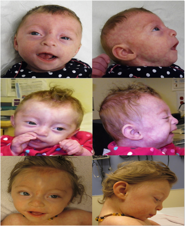
(left to right, top to bottom) Clinical photographs of the patient at 3, 6, and 14 months of age. Dysmorphic features shown here include hypertelorism, shallow orbits, smooth philtrum, micrognathia, fleshy ears, hypoplastic nipples and cutis marmorata.
Unfortunately, not all stories have a happy ending. Three of the documented cases involved babies who did not survive, passing away due to severe skeletal dysplasia in utero. Yet, others show resilience—one 10-year-old patient is managing her condition with fewer hospital visits despite continued developmental challenges.
Genetics is like a complex code, and in ALG9-CDG, scientists have identified three key mutations that disrupt this code:
p.(Tyr287Cys)
p.(Glu530Lys)
c.(1173 +2T > A)
These mutations don’t all act the same way. Some, such as splicing mutations, can lead to more severe forms of the disease, whereas missense mutations—which change a single amino acid in a protein—tend to result in a less intense disease progression. Researchers are still piecing together the puzzle, but these findings offer hope for better understanding how different mutations impact the severity of symptoms.
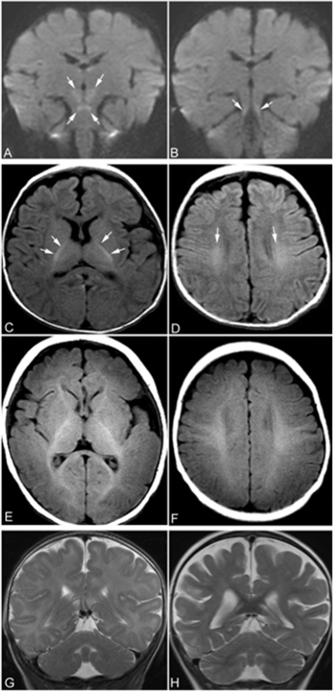
If we could peek inside the brain of an ALG9-CDG patient, what would we see? Brain imaging has revealed some telling clues. Patients often experience delayed myelination, meaning the protective coating around their brain's nerve fibers doesn’t develop as quickly as it should. This can lead to cerebral atrophy, or shrinking of brain tissue. As the disease progresses, follow-up MRIs show that certain regions of the brain may also exhibit restricted diffusion, a signal that something deeper is at play—perhaps even cytotoxic edema, where brain cells begin to swell.
These neuroimaging results add another layer to understanding ALG9-CDG, helping doctors better monitor the disease over time.
ALG9-CDG, like many rare disorders, is a field where knowledge is still growing. Each new case offers more insight into how this condition presents and progresses, and every new genetic discovery brings us closer to understanding how these mutations impact the body. Research into this condition isn’t just about documenting symptoms—it’s about finding ways to improve outcomes. As more cases are studied, there’s hope that better diagnostic tools and treatment strategies will emerge. For now, families and patients with ALG9-CDG hold on to the possibility of a future where more answers lead to improved care.
ALG9-CDG may be a rare condition, but its story is one of resilience, scientific curiosity, and the determination to unlock its secrets. The more we learn about this complex disorder, the closer we come to improving the lives of those affected. For now, every new discovery is a step forward, bringing hope to families navigating the challenges of ALG9-CDG.
Stay tuned for more breakthroughs as science continues to unravel the mysteries behind this rare but important condition.
With every rare disease, there’s a human story behind the science—a journey that demands attention, empathy, and relentless pursuit of answers. ALG9-CDG may be one puzzle among many, but it’s a puzzle we’re determined to solve.
Research Assistant, exRNA Therapeutics
Hypertension and brachydactyly syndrome (HTNB), also known as Bilginturan syndrome, is a rare genetic condition that presents a unique combination of symptoms, including brachydactyly type E and severe, salt-independent hypertension. This blog aims to explore the genetic underpinnings, clinical implications, and recent findings related to this intriguing syndrome.
HTNB is characterized by:
Without treatment, individuals with HNTB face a high risk of stroke before the age of 50.

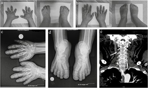
Clinical features of this HTNB family. (a) Photographs of brachydactyly in the hands and feet of the proband. (b) Photographs of the normal hands and feet of the probands’ mother. (c) An X-ray of the proband's short hands indicating involvement in all metacarpals and phalanges. (d) An X-ray of the proband’s feet showing short metatarsal bones. (e) A computed tomography image showing a stenotic right vertebral artery (white arrow) in the proband.
The genetic basis of HNTB was initially linked to chromosome 12p12.2-p11.2. In 2015, significant progress was made when researchers identified mutations in the PDE3A gene through whole-genome sequencing in Turkish families affected by the syndrome.
PDE3A Gene: This gene encodes for the enzyme phosphodiesterase 3A, which regulates important physiological processes by breaking down cyclic AMP (cAMP) and cyclic GMP. Mutations in PDE3A lead to an upregulation of cAMP-hydrolytic activity, resulting in decreased cAMP levels in vascular smooth muscle cells. This reduction may trigger arteriogenesis and contraction, ultimately causing hypertension and increasing the risk of stroke.
In a groundbreaking study, researchers reported a new gain-of-function mutation (c.1346G>A) in PDE3A within a Chinese family with HTNB. This mutation is located within the cluster of previously identified mutations and further supports the hypothesis that alterations in PDE3A are central to the pathogenesis of this syndrome.
The identification of this mutation not only deepens our understanding of HTNB but also highlights potential avenues for therapeutic intervention. By targeting the pathways influenced by PDE3A mutations, there may be opportunities to develop treatments that can mitigate hypertension and its associated risks.
HTNB is a complex genetic disorder with significant implications for affected individuals. As research continues to unravel the intricacies of this syndrome, including its genetic basis and clinical manifestations, it paves the way for improved diagnostic methods and potential therapeutic strategies. The recent discovery of new mutations in PDE3A underscores the importance of ongoing research in understanding rare diseases like HTNB and their impact on patient health.
Research Assistant, exRNA Therapeutics
The phosphodiesterase 3A (PDE3A) gene encodes an enzyme responsible for hydrolyzing cyclic adenosine monophosphate (cAMP), a vital second messenger involved in various physiological processes. By regulating cAMP levels, PDE3A plays a significant role in controlling vascular tone and cardiac function.
Regulation of cAMP Levels: PDE3A terminates cAMP signaling, influencing smooth muscle contraction and relaxation.
Isoforms: The gene produces three main isoforms—PDE3A1, PDE3A2, and PDE3A3—each with distinct roles in different tissues.
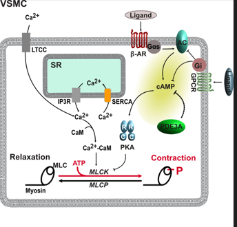
In VSMCs, cAMP signaling is crucial for regulating contraction and relaxation. When activated by G-protein-coupled receptors (GPCRs), cAMP levels rise, leading to the activation of protein kinase A (PKA). PKA phosphorylates various proteins that inhibit myosin light chain kinase (MLCK), thereby promoting relaxation:
FIGURE1:-cAMP Regulation in Vascular Smooth Muscle Cells
Mutations in the PDE3A gene are linked to a rare form of hypertension known as hypertension with brachydactyly (HTNB). These mutations result in hyperactive PDE3A enzymes that disrupt normal cAMP signaling. This disruption leads to increased vascular smooth muscle cell proliferation and compromised vasodilation, resulting in severe hypertension without the typical cardiac damage.
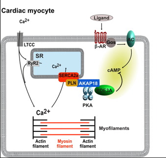
In cardiac myocytes, cAMP signaling also plays a crucial role in contractility. Similar to VSMCs, elevated cAMP levels enhance contractile function by modulating calcium handling:
Figure 2: cAMP Regulation in Cardiac Myocytes emphasizing its role in contractility.
The interplay between the PDE3A gene and cAMP signaling compartments is vital for maintaining vascular and cardiac health. Understanding how mutations in PDE3A lead to hypertension while protecting against cardiac damage opens new avenues for research into therapeutic strategies targeting these pathways. By focusing on the molecular mechanisms underlying these processes, we can better comprehend blood pressure regulation and develop innovative treatments for hypertension-related conditions.
Research Assistant, exRNA Therapeutics
Jervell and Lange-Nielsen Syndrome (JLNS) is a rare but critical genetic disorder that intertwines auditory and cardiac health, primarily caused by mutations in the KCNE1 and KCNQ1 genes. Understanding these genes’ functions provides insight into how they contribute to the syndrome's symptoms, which include profound hearing loss and an increased risk of life-threatening cardiac arrhythmias.
KCNE1 encodes a regulatory subunit of the potassium channel complex formed with KCNQ1, which is essential for normal cardiac repolarization and auditory function. Mutations in these genes lead to two types of JLNS:
Both types are inherited in an autosomal recessive manner, meaning that both copies of the gene must be mutated for the individual to exhibit symptoms. This genetic interplay highlights the critical roles these proteins play in both hearing and heart function.
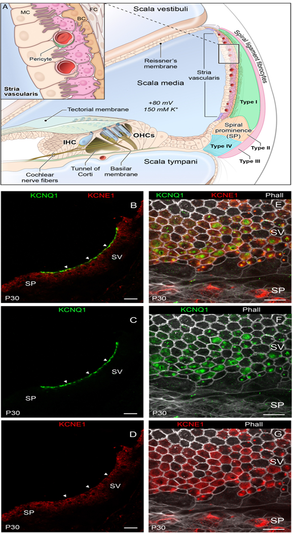
The inner ear's stria vascularis is crucial for maintaining the ionic environment necessary for hearing. Both KCNE1 and KCNQ1 are integral to creating the endocochlear potential, which is vital for hair cell function—the cells responsible for converting sound vibrations into neural signals. When either gene is mutated, it disrupts this process, leading to congenital profound sensorineural deafness.

Individuals with JLNS face significant cardiac risks due to prolonged QT intervals, which can lead to dangerous arrhythmias. The QT interval represents the time it takes for the heart's ventricles to depolarize and repolarize. Prolongation can result in syncopal episodes or even sudden cardiac death, particularly during physical exertion or emotional stress.
The clinical presentation of JLNS typically includes:
Given the serious implications of these symptoms, early diagnosis through genetic testing is crucial. Families with a history of congenital deafness or unexplained cardiac events should consider genetic counseling.
Genetic testing can identify mutations in KCNE1 and KCNQ1, providing valuable information for management and family planning. Individuals identified as carriers may not exhibit symptoms but can pass on the mutations to their offspring.
Jervell and Lange-Nielsen Syndrome serves as a poignant reminder of how interconnected our genetic makeup is with our health outcomes. The roles of KCNE1 and KCNQ1 in both hearing and cardiac function underscore the need for comprehensive genetic testing and counseling. As research continues to advance our understanding of these genes, it opens doors for improved management strategies for individuals affected by this complex syndrome. By delving into the genetics of JLNS, we not only illuminate the mechanisms behind hearing loss and cardiac issues but also emphasize the importance of awareness and early intervention in managing this multifaceted disorder.
Research Assistant, exRNA Therapeutics
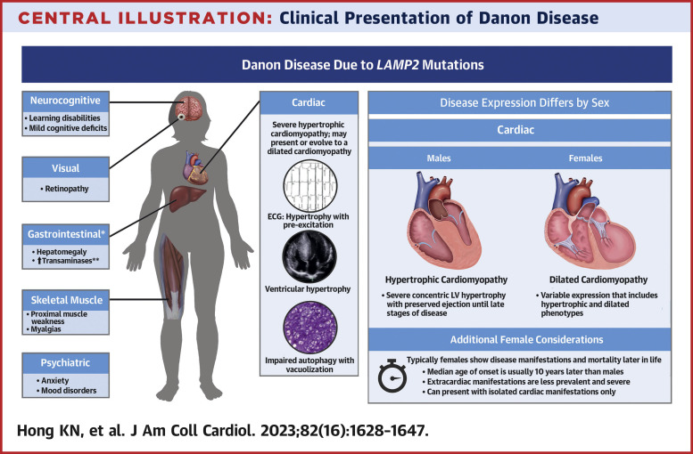
Danon disease is an X-linked dominant disorder caused by mutations in the LAMP2 gene, which encodes a protein essential for lysosomal function. Initially thought to be a lysosomal storage disease due to similarities with Pompe disease, it was reclassified after the genetic basis was identified in 2000. The absence of LAMP2 protein disrupts cellular processes, leading to the accumulation of autophagic materials and glycogen in muscle cells.
Danon disease typically presents with a triad of symptoms:
If you or someone you know has been diagnosed with Danon disease, consider sharing your story with us. Your experiences can help raise awareness and support others facing similar challenges.
Diagnosing Danon disease involves a combination of clinical evaluation and genetic testing:

The prevalence of Danon disease is not well established but may be increasing due to improved genetic testing availability. Early detection can significantly impact management and outcomes.
While there is no cure for Danon disease, management focuses on alleviating symptoms and preventing complications:
Research is ongoing to better understand the molecular mechanisms behind Danon disease and explore potential therapies. Engaging with clinical trials could provide opportunities for affected individuals to contribute to advancements in treatment.
Danon disease is a multifaceted condition that requires comprehensive management tailored to individual needs. By raising awareness and fostering community support, we can improve outcomes for those affected by this rare disorder.
Research Assistant, exRNA Therapeutics
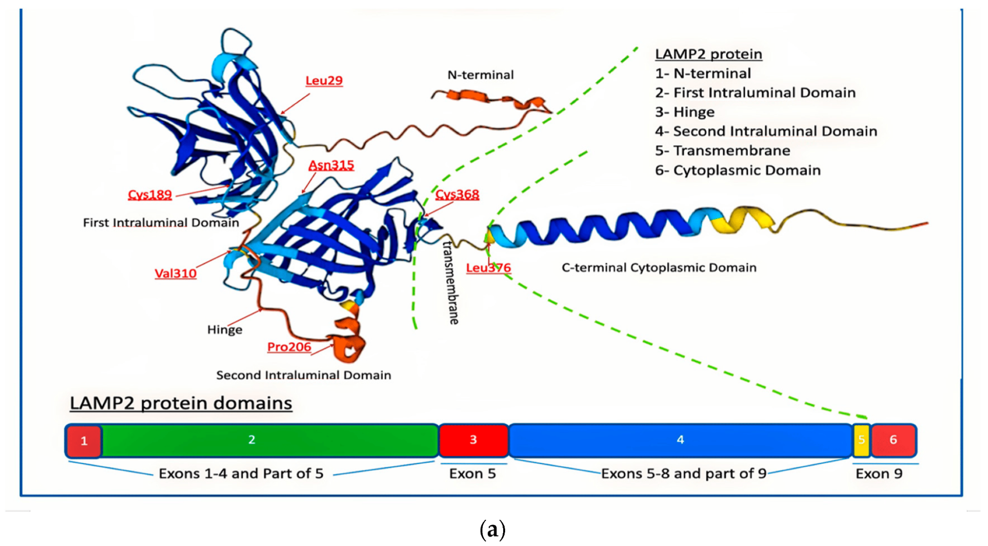
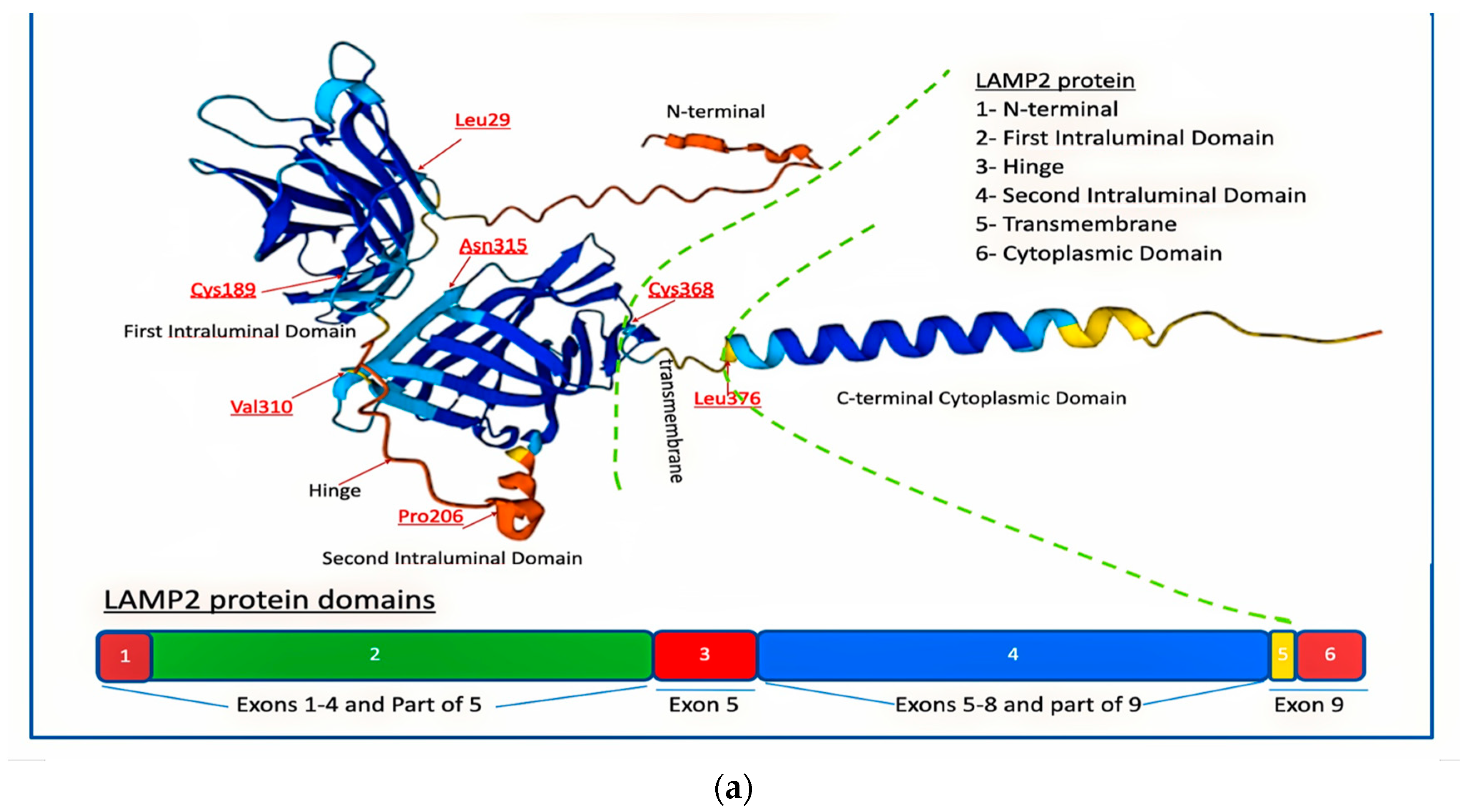
Lysosome-associated membrane protein 2 (LAMP2) plays a crucial role in cellular homeostasis, particularly in lysosomal function. It is a transmembrane protein found primarily in lysosomes, the organelles responsible for breaking down waste materials within the cell. When mutations affect LAMP2, it disrupts cellular processes, leading to diseases such as Danon disease, a rare and severe genetic disorder. Let's dive deeper into the structure and function of LAMP2, as illustrated in the diagram, and how its mutations are connected to disease development.
The structure of LAMP2 is composed of multiple distinct domains, each performing critical functions:

This is the starting point of the protein and is extracellular, meaning it extends outside the lysosome. The N-terminal helps regulate interactions between the lysosome and other parts of the cell.
The first intraluminal domain lies inside the lysosome, with crucial amino acid residues such as Cys189, Leu29, and Asn315 highlighted. These amino acids are essential for the stability and function of the protein.
This region acts as a flexible link between different parts of the protein, allowing it to change shape and adjust based on cellular needs. Val310 and Pro206 are found in this section and contribute to the flexibility of the protein.
Similar to the first intraluminal domain, this region is critical for the lysosomal function. It contains important residues like Cys368, which help maintain structural integrity.
This region anchors LAMP2 in the lysosomal membrane, providing the structural support to keep it embedded in the lysosome wall. Mutations here can destabilize the protein’s attachment to the lysosome.
The C-terminal tail extends into the cytoplasm, influencing various signaling pathways that govern lysosomal function. The Leu376 residue, located here, is vital for these cellular signals.
As illustrated in the lower section of the diagram, LAMP2 is made up of several exons (segments of genes coding for parts of the protein), each contributing to different protein domains:
Danon disease is caused by mutations in the LAMP2 gene. These mutations result in either a complete or partial loss of LAMP2 protein function, disrupting the lysosome's ability to degrade cellular waste. This leads to the accumulation of materials in cells, particularly affecting the heart, muscles, and brain. Symptoms of Danon disease include:
LAMP2 is not only critical for cellular waste management but also plays a role in other cellular processes such as autophagy, a mechanism where cells digest their damaged or unnecessary components. Proper autophagy helps cells survive under stress by recycling resources. Without functional LAMP2, autophagic pathways are impaired, contributing to the accumulation of toxic materials and ultimately to cell death in affected tissues.
LAMP2 is a vital player in maintaining cellular health, and mutations affecting this protein lead to the onset of Danon disease. Understanding its structure, especially the role of key domains and exons, helps researchers and clinicians develop better diagnostic and therapeutic strategies for managing lysosomal storage diseases.
Stay tuned for further insights into genetic disorders and how cutting-edge research is unlocking new treatments for conditions like Danon disease.
Research Assistant, exRNA Therapeutics
Brugada Syndrome (BrS) is a rare inherited arrhythmia disorder that significantly increases the risk of sudden cardiac death, particularly in young adults. This blog will explore the genetic factors associated with Brugada Syndrome, focusing on specific genes such as SCN3B, CACNA1C, and CACNB2, and how they contribute to different types of BrS.
Brugada Syndrome is characterized by a distinctive electrocardiographic (ECG) pattern and is often asymptomatic until it results in severe complications like syncope or sudden cardiac arrest. The syndrome is classified into three main types based on the ECG findings:
BrS has been linked to mutations in various genes that affect cardiac ion channels, leading to impaired electrical conduction and increased arrhythmia risk. The following genes are particularly significant:

The SCN3B gene encodes the beta subunit of the sodium channel Navβ3. Mutations in this gene are rare but can lead to reduced sodium current (INa), contributing to the pathophysiology of BrS. Although SCN5A mutations are more common, SCN3B variants highlight the genetic complexity of the syndrome.
The CACNA1C gene encodes the alpha subunit of the Cav1.2 calcium channel, while CACNB2 encodes its beta subunit. Both genes play essential roles in calcium influx during cardiac action potentials. Mutations can lead to decreased calcium current (ICa), further complicating cardiac electrical activity and increasing arrhythmia risk.
Two primary hypotheses explain the mechanisms behind BrS
Understanding the genetic basis of BrS can significantly impact diagnosis, risk stratification, and treatment strategies. Genetic testing can help identify at-risk individuals, especially those with a family history of sudden cardiac death.
Brugada Syndrome represents a complex interplay between genetic factors and clinical manifestations. The identification of genes like SCN3B, CACNA1C, and CACNB2 adds depth to our understanding of this potentially fatal condition. Ongoing research into these genetic factors is crucial for developing personalized treatment strategies and improving patient outcomes.
Research Assistant, exRNA Therapeutics
Timothy Syndrome, also known as Long QT Syndrome Type 8 (LQT8), is characterized by a range of symptoms affecting the heart, skeleton, metabolic system, and brain. The most notable cardiac manifestation is the prolongation of the QT interval, which can lead to serious arrhythmias and even cardiac arrest at a young age.
Timothy Syndrome (TS) is associated with multisystem dysfunction and developmental defects:


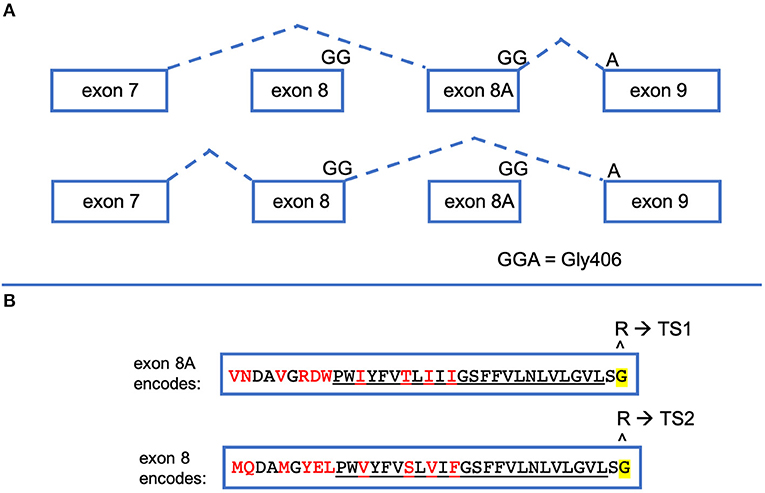
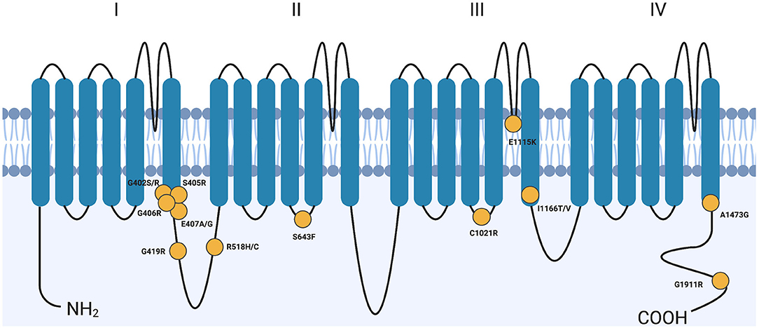
Timothy Syndrome is primarily caused by mutations in the CACNA1C gene, which encodes the α1C subunit of voltage-gated calcium channels. These channels are essential for calcium ion flow into cells, influencing numerous physiological functions. The most common mutation linked to TS is Gly406Arg, which can occur in either exon 8 or exon 8A of the CACNA1C gene.
Type 1 (TS1): Caused by mutations in exon 8A, often resulting in more severe symptoms including syndactyly and neurodevelopmental delays.
Type 2 (TS2): Linked to mutations in exon 8, typically presenting with milder symptoms and lacking syndactyly but may include hip dysplasia.
Mutations in CACNA1C lead to biophysical changes in the calcium channels, often resulting in a gain-of-function phenotype. This means that the mutated channels open more readily or remain open longer than normal, causing excessive calcium influx into cells. This dysregulation can lead to:
While there is no cure for Timothy Syndrome, several treatment strategies aim to manage symptoms:
Timothy Syndrome presents a complex interplay of genetic mutations and clinical manifestations that require careful management. Ongoing research continues to uncover the nuances of this disorder, paving the way for better diagnostic tools and treatment options. As awareness grows and more cases are identified, it is crucial for patients and families to stay informed about available resources and support networks dedicated to managing this rare condition.
© eXRna Rare. All Rights Reserved 2024.
Designed by eXRna Rare
Distributed by eXRna Rare