Research Assistant, exRNA Therapeutics
Kartagener’s Syndrome is a rare hereditary genetic disease (1: 20,000 cases approx) and occurs from birth. Its inheritance is autosomal recessive. This syndrome is also known as primary ciliary dyskinesia (PCD) syndrome. It’s characterized by causing alterations in the cilia structure and function present in the respiratory system cells and gonadal tissues (fallopian tubes and sperm). These alterations affect the coordinated and normal ciliary movement. This lack of movement in the cilia causes recurrent infections of the upper and lower respiratory tract and infertility.
Patients with PCD are characterized by presenting:
Chronic sinusitis
Bronchiectasis
Situs inversus ( in this the heart shifted to right side)
As it is a genetic disease, the cause of Kartagener syndrome is a mutation in different genes associated with the alteration of the cilia. Although DNAH 11 a gene encode a protein that is a part of the outer dynein arm of cilia and is required for cilia motility and CCDC40, responsible for assembly of internal dynein arm.Mutations in these genes cause the loss of the dynein arms of the cilia and thus prevent their movement.
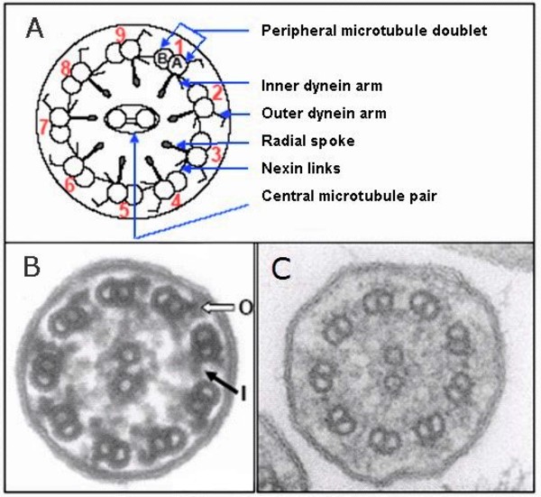
A ) schematic diagram of a cilium; B ) electron microscopic image of a normal cilium with "O" representing the outer dynein arm and "I" representing the inner dynein arm; C) electron microscopic image of a cilium of an individual with primary ciliary dyskinesia showing absence of dynein arms
This syndrome has a negative effect on fertility, both in men and women.
Specifically, in males there’s an alteration of sperm motility, due to abnormalities in the structure of the sperm’s tail (flagellum). This structural alteration generates an absence of motility in the sperm cells (asthenozoospermia), which prevents them from advancing through the female genital tract and fertilizing the ovum.
In case of women, the cilia found in the fallopian tubes are affected, its function is to help the ovum to move to the fertilization area, thus preventing the ovum or embryo from advancing through the tube. In women affected by this syndrome, the risk of ectopic pregnancy is higher
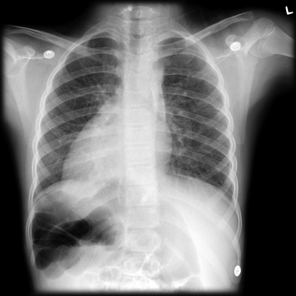
Imaging- X-ray showing situs inversus. Note the liver and heart on opposite sides of the body
Research Assistant, exRNA Therapeutics
Catamenial Pneumothorax (CP) is an extremely rare, but very dangerous condition. While it only comprises 3%-6% of all spontaneous pneumothorax cases among menstruating women.Recurrent spontaneous pneumothorax associated with the menstrual cycle was described first by Maurer et al in 1958. The term catamenial pneumothorax was established by Lillington et al in 1972. The main criteria for the diagnosis of catamenial pneumothorax are known as the sine qua non (without which not) criteria, which is spontaneous pneumothorax within 72 hours before or after the start of a menstrual period. In addition to this, CP often presents with concomitant endometriosis, right-sided location, and pleural lesions. There are following four postulates that somehow explain the CP:-

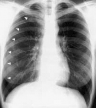
Figure 1 Posteroanterior chest radiograph reveals right-sided pneumothorax (arrows indicate edge of visceral pleura).
In summary, our report demonstrates a case of catamenial pneumothorax in a patient with endometriosis. The occurrence of this condition is very rare and only comprises a very small amount of all spontaneous pneumothorax cases in young women.
Research Assistant, exRNA Therapeutics
Liver cirrhosis is a progressive liver disease characterized by extensive fibrosis, impaired liver function, and significant hormonal disturbances. The SMAD4 gene, a critical mediator in the TGF-β (Transforming Growth Factor-beta) signaling pathway, plays a key role in the progression of fibrosis and the maintenance of cellular homeostasis. This report explores how SMAD4, through its involvement in liver fibrosis and systemic inflammation, can indirectly affect fertility in individuals with liver cirrhosis, contributing to hormonal imbalances, reproductive tissue fibrosis, and reduced fertility in both men and women.
One of the major consequences of liver cirrhosis is hormonal dysregulation, which affects fertility in both men and women:
The liver plays a key role in metabolizing estrogen and producing sex hormone-binding globulin (SHBG). In cirrhosis, reduced liver function leads to decreased estrogen metabolism and lower levels of SHBG, resulting in elevated circulating estrogen (Dulai et al., 2016). This excess estrogen is associated with hypogonadism in men and menstrual irregularities in women.
Elevated estrogen levels lead to gynecomastia, reduced libido, erectile dysfunction, and impaired spermatogenesis due to low testosterone. Testicular atrophy and reduced sperm count are common in men with cirrhosis
In women, liver cirrhosis can result in anovulation, irregular menstrual cycles, and premature ovarian failure due to the hormonal imbalance caused by disrupted liver function (Selvaggi et al., 2018). The buildup of estrogen in the body can interfere with normal ovarian and uterine function, reducing the chances of conception.
Apart from its role in liver fibrosis, SMAD4 is also implicated in the fibrotic processes in other organs, including the reproductive system. The TGF-β/SMAD4 signaling pathway is involved in the fibrosis of reproductive tissues, which can contribute to infertility
Overactivation of the TGF-β pathway, driven by SMAD4, can promote ovarian and uterine fibrosis. Fibrosis in these tissues can interfere with normal ovarian function, ovulation, and embryo implantation, all of which are critical for fertility.
Similar fibrotic mechanisms can affect testicular function, leading to testicular fibrosis, impaired spermatogenesis, and reduced testosterone production, all of which contribute to infertility in men with cirrhosis
Liver cirrhosis induces a state of chronic systemic inflammation and oxidative stress, which further disrupts reproductive health. SMAD4, through the TGF-β pathway, is a key regulator of the inflammatory response:
Chronic inflammation can lead to testicular damage, reduced sperm quality, and increased oxidative stress, all of which are detrimental to fertility
inflammatory cytokines can impair ovarian function, disrupt the menstrual cycle, and affect egg quality, reducing fertility
While SMAD4 does not directly control reproductive hormones, its role in liver fibrosis, reproductive tissue fibrosis, and systemic inflammation indirectly contributes to infertility in individuals with liver cirrhosis. The impaired liver function due to cirrhosis leads to significant hormonal imbalances, particularly elevated estrogen levels in both men and women. SMAD4-driven fibrosis in reproductive organs can further exacerbate these effects, leading to testicular and ovarian dysfunction, reduced sperm production, and menstrual irregularities. Understanding these pathways highlights the interconnected nature of liver disease and reproductive health.
Research Assistant, exRNA Therapeutics
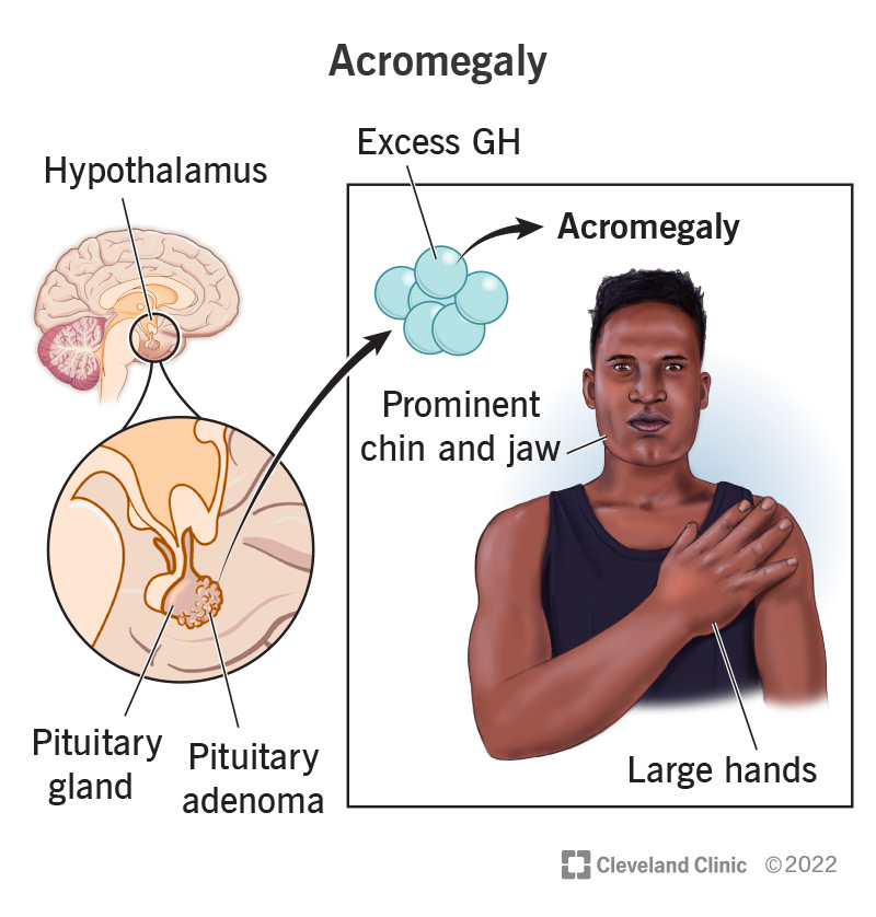
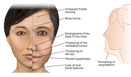
Acromegaly is a rare endocrine disorder characterized by excessive production of growth hormone (GH), typically due to a benign tumor called a pituitary adenoma. Acromegaly typically manifests in adults, while gigantism occurs in children before their growth plates close. The prevalence of acromegaly is approximately 40-130 cases per million people, with about 3-4 new cases per million each year.Symptoms of acromegaly include enlarged hands and feet, coarsened facial features (such as a protruding jaw and enlarged nose), joint pain, and potential complications like cardiovascular disease, diabetes, and joint problems, making early diagnosis crucial. The condition is primarily caused by pituitary adenomas, which lead to excessive production of GH.The overproduction of GH leads to increased levels of insulin-like growth factor 1 (IGF-1), which drives the abnormal growth of bones and tissues. In some cases, genetic mutations, such as in the AIP (aryl hydrocarbon receptor-interacting protein) gene, are implicated, particularly in familial forms. Other genes like GNAS, which causes McCune-Albright syndrome, may also be associated. Other genetic predispositions may occur due to familial conditions such as Multiple Endocrine Neoplasia type 1 (MEN1), where tumors can develop in multiple endocrine glands.Treatment primarily focuses on reducing GH levels and tumor size. Surgical removal of the adenoma is often the first line of treatment, with transsphenoidal surgery being the most common approach. For individuals whose tumors are not completely removed or are not surgical candidates, medical therapies like somatostatin analogs and growth hormone receptor antagonists can effectively manage symptoms and hormone levels. In some cases, radiation therapy may be used to control tumor growth and hormone secretion.Early diagnosis and treatment are crucial to mitigate the serious health risks associated with this disorder.


© eXRna Rare. All Rights Reserved 2024.
Designed by eXRna Rare
Distributed by eXRna Rare