Research Assistant (exRNA Therapeutics)
Autosomal recessive spondylocostal dysostosis (ARSD) is a rare congenital disorder characterized by abnormal vertebral and rib development, leading to a shortened trunk, scoliosis, and other skeletal malformations. The condition arises from mutations in genes responsible for the proper formation of the axial skeleton, specifically during embryonic development. ARSD is a severe form of skeletal dysplasia, a group of disorders marked by abnormal bone and cartilage growth.
Most cases of ARSD are linked to mutations in genes such as DLL3, MESP2, LFNG, and HES7, which are integral to the Notch signaling pathway. This pathway governs segmentation of the vertebrae and ribs, and any disruption leads to the characteristic malformations seen in ARSD. Patients typically exhibit a unique pattern of vertebral segmentation, with fused or absent ribs and malformed vertebrae. The severity of the condition varies, and while some patients experience severe respiratory complications due to rib cage deformities, others may live relatively normal lives with minimal intervention.
Given the rare and complex nature of ARSD, traditional treatment options are limited, focusing mainly on symptom management through physical therapy, bracing, or surgical correction of spinal deformities. However, recent advancements in genetic research have paved the way for innovative therapies that address the underlying genetic defects. One such promising approach is the use of antisense oligonucleotide (ASO) therapy.
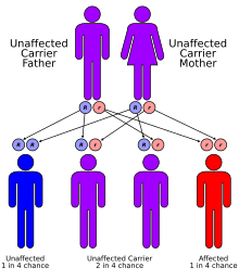
ARSD is inherited in an autosomal recessive pattern, meaning both copies of the mutated gene must be inherited from each parent for the disease to manifest. This makes the condition particularly rare, as both parents must carry the defective gene without necessarily displaying symptoms themselves. As a result, ARSD is most commonly observed in families with a history of consanguinity.
The genes implicated in ARSD, such as DLL3 and MESP2, are crucial to a highly regulated developmental process known as somitogenesis. This process ensures the precise formation of somites, which are structures that give rise to the vertebrae and ribs. Mutations in these genes disrupt this process, leading to vertebral segmentation anomalies.
Research into ARSD has been challenging due to its rarity and the complexity of the genetic mechanisms involved. Advances in nextgeneration sequencing technologies have enabled more precise identification of mutations, leading to a better understanding of the molecular underpinnings of the disease. Despite these advances, the development of therapies targeting the genetic root of ARSD has remained elusive.
One of the most promising developments in the treatment of genetic disorders is the emergence of antisense oligonucleotide (ASO) therapy. ASOs are short, synthetic strands of nucleotides designed to bind to specific RNA sequences, effectively modulating gene expression. By binding to the RNA transcript, ASOs can either prevent the production of a harmful protein or restore the function of a defective one.
In the context of ARSD, ASO therapy could target the aberrant RNA transcripts produced by the mutated genes, potentially correcting the downstream effects of these mutations. The therapy works by altering the splicing of premRNA or by inducing the degradation of the mutated transcript, thus preventing the synthesis of dysfunctional proteins that disrupt skeletal development.
The potential for ASO therapy to treat ARSD lies in its ability to precisely target the genetic mutations responsible for the disease. For example, a mutation in the DLL3 gene could be addressed by designing an ASO that binds to the mutated transcript, thereby reducing the production of the defective protein and allowing for more normal skeletal development. This approach has already shown promise in other rare genetic disorders, such as spinal muscular atrophy (SMA), where ASO therapy has been successfully implemented.
While the potential for ASO therapy in ARSD is significant, several challenges remain. The first challenge lies in the delivery of ASOs to the affected tissues. Effective delivery of ASOs to the developing skeletal system is critical, as ARSD primarily affects the bones and cartilage. Achieving targeted delivery to these tissues, especially during the critical stages of embryonic development, presents a significant hurdle in translating ASO therapy from theory to practice.
Another challenge is the precise identification of the mutation to be targeted. Given the genetic heterogeneity of ARSD, with multiple different mutations in various genes leading to the disorder, a onesizefitsall approach is unlikely to be effective. Instead, personalized therapies must be developed to target the specific mutation present in each individual patient.
Moreover, longterm safety and efficacy studies are required to ensure that ASO therapy does not produce unintended side effects, such as immune reactions or offtarget effects on other genes. While ASOs are generally welltolerated, their use in a developing embryo raises unique concerns that must be addressed through rigorous clinical testing.
Despite these challenges, the future of ASO therapy in treating ARSD is promising. As our understanding of the genetic basis of the disease deepens and delivery methods improve, the potential for ASO therapy to correct the underlying genetic defects in ARSD becomes more tangible. Ongoing research is focused on optimizing ASO design, enhancing tissuespecific delivery, and ensuring longterm safety and efficacy.
Additionally, the use of patientderived stem cells and geneediting technologies such as CRISPR may further complement ASO therapy, offering a multipronged approach to tackling ARSD at its genetic roots. With continued investment in research and collaboration between academia, industry, and patient advocacy groups, ASO therapy could one day transform the lives of patients with ARSD and other rare genetic disorders.
Autosomal recessive spondylocostal dysostosis represents a rare but devastating genetic condition that disrupts normal skeletal development. While current treatments focus on managing the symptoms, recent advances in genetic research have opened the door to novel therapies, such as antisense oligonucleotides, that aim to correct the underlying genetic mutations. Though challenges remain, the potential of ASO therapy to provide a targeted, personalized approach to ARSD treatment is an exciting development in the field of rare disease research.
As researchers continue to unlock the mysteries of ARSD and refine ASO therapies, the hope for effective treatments that can significantly improve the quality of life for patients becomes more attainable. The journey from genetic discovery to therapeutic innovation is a complex one, but with perseverance and collaboration, the future of ARSD treatment looks bright.
Research Assistant (exRNA Therapeutics)
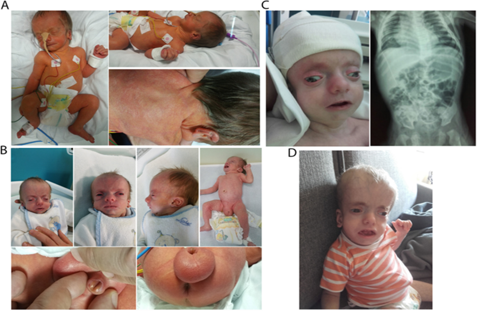
Boomerang Dysplasia is an exceptionally rare and severe genetic disorder that primarily affects skeletal development. This condition is characterized by profound skeletal abnormalities that can be detected as early as during prenatal development. The term "boomerang" refers to the distinctive shape of the bones in the limbs, which take on a boomerang like appearance in imaging studies, particularly in the arms and legs. Infants born with Boomerang Dysplasia typically face multiple, life threatening complications, and unfortunately, survival beyond birth is exceedingly rare.
Though incredibly rare, the scientific community has made significant strides in identifying the genetic underpinnings of this disorder. This understanding has opened the door to potential novel therapeutic interventions, including antisense oligonucleotide (ASO) therapy, a promising approach targeting the genetic basis of Boomerang Dysplasia.
Boomerang Dysplasia is caused by mutations in the FLNB gene (Filamin B). This gene encodes a protein that is essential for normal skeletal development, as it plays a pivotal role in the organization of the actin cytoskeleton in developing cells. Actin filaments are crucial for maintaining cellular shape and providing the structural integrity necessary for tissues like bone to develop correctly.
Mutations in the FLNB gene disrupt the formation of filamin B, resulting in the abnormal development of the skeletal system. This leads to the hallmark features of Boomerang Dysplasia, including:
The disease is typically diagnosed through prenatal ultrasound and genetic testing. However, due to the severity of the condition, infants born with Boomerang Dysplasia rarely survive, often succumbing to complications such as respiratory failure due to underdeveloped lungs or severe skeletal malformations.
To date, there is no cure for Boomerang Dysplasia, and treatment options are extremely limited due to the lifethreatening nature of the disorder. Medical management primarily involves palliative care to ensure comfort and address symptoms. Traditional therapeutic approaches, such as surgery or drug treatments, are often not feasible due to the extent of the skeletal abnormalities and the rapid progression of the disease.
However, advances in genebased therapies, particularly antisense oligonucleotides (ASOs), have raised the possibility of treating the underlying genetic cause of Boomerang Dysplasia rather than managing symptoms alone.
Antisense oligonucleotides (ASOs) are short, synthetic strands of nucleic acids designed to bind to specific RNA sequences. By targeting and binding to the mutated RNA transcript responsible for the production of dysfunctional proteins. This approach is particularly promising for monogenic diseases like Boomerang Dysplasia, where a single mutation in the FLNB gene is responsible for the disorder.
1. Targeting Mutant RNA: ASOs are designed to bind to the mutated RNA, preventing it from being translated into a defective protein or inducing its degradation before translation.
2 .Precision Medicine: ASOs offer a precision medicine approach, allowing scientists to tailor treatment to the specific mutation found in a patient’s genetic code.
In the context of Boomerang Dysplasia, ASO therapy holds the potential to directly target the FLNB gene mutations that lead to the improper formation of skeletal structures. By modifying the expression of the mutated FLNB gene, ASOs could theoretically prevent the development of the skeletal malformations that define this disorder.
Several features make ASO therapy a compelling option for Boomerang Dysplasia:
1. Direct Gene Targeting: ASOs offer the potential to intervene at the RNA level, directly targeting the root cause of Boomerang Dysplasia rather than simply managing its symptoms.
2. Potential for Early Intervention: Given that Boomerang Dysplasia’s skeletal abnormalities manifest early in fetal development, ASO therapy could potentially be administered during pregnancy or shortly after birth, offering a chance to intervene before the disease progresses too far.
3. Customizability: Since ASOs can be designed to target specific mutations in the FLNB gene, this approach allows for a highly personalized treatment plan that could accommodate individual genetic variations within the disorder.
4. NonInvasive Treatment: ASO therapies are typically administered via injection, offering a less invasive alternative to surgical interventions or other more intensive treatments.
Despite its promise, there are still challenges to overcome before ASO therapy can become a viable treatment for Boomerang Dysplasia:
The emergence of antisense oligonucleotide therapy is a significant breakthrough in the treatment of rare genetic diseases, including Boomerang Dysplasia. By targeting the underlying genetic mutations responsible for these conditions, ASO therapy represents a shift towards precision medicine that goes beyond symptom management to potentially curing or mitigating the disease at its source.
While more research is needed to address the challenges associated with Boomerang Dysplasia, the promise of ASO therapy offers hope to affected families. At your organization, we are committed to advancing the development of genetargeted therapies like ASOs, with the ultimate goal of providing effective treatments for rare and devastating conditions like Boomerang Dysplasia.
Research Assistant (exRNA Therapeutics)
Boomerang Dysplasia is an exceptionally rare and severe genetic disorder that primarily affects skeletal development. This condition is characterized by profound skeletal abnormalities that can be detected as early as during prenatal development. The term "boomerang" refers to the distinctive shape of the bones in the limbs, which take on a boomerang like appearance in imaging studies, particularly in the arms and legs. Infants born with Boomerang Dysplasia typically face multiple, life threatening complications, and unfortunately, survival beyond birth is exceedingly rare.
Though incredibly rare, the scientific community has made significant strides in identifying the genetic underpinnings of this disorder. This understanding has opened the door to potential novel therapeutic interventions, including antisense oligonucleotide (ASO) therapy, a promising approach targeting the genetic basis of Boomerang Dysplasia.
Boomerang Dysplasia is caused by mutations in the FLNB gene (Filamin B). This gene encodes a protein that is essential for normal skeletal development, as it plays a pivotal role in the organization of the actin cytoskeleton in developing cells. Actin filaments are crucial for maintaining cellular shape and providing the structural integrity necessary for tissues like bone to develop correctly.
Mutations in the FLNB gene disrupt the formation of filamin B, resulting in the abnormal development of the skeletal system. This leads to the hallmark features of Boomerang Dysplasia, including:
The disease is typically diagnosed through prenatal ultrasound and genetic testing. However, due to the severity of the condition, infants born with Boomerang Dysplasia rarely survive, often succumbing to complications such as respiratory failure due to underdeveloped lungs or severe skeletal malformations.
To date, there is no cure for Boomerang Dysplasia, and treatment options are extremely limited due to the lifethreatening nature of the disorder. Medical management primarily involves palliative care to ensure comfort and address symptoms. Traditional therapeutic approaches, such as surgery or drug treatments, are often not feasible due to the extent of the skeletal abnormalities and the rapid progression of the disease.
However, advances in genebased therapies, particularly antisense oligonucleotides (ASOs), have raised the possibility of treating the underlying genetic cause of Boomerang Dysplasia rather than managing symptoms alone.
Antisense oligonucleotides (ASOs) are short, synthetic strands of nucleic acids designed to bind to specific RNA sequences. By targeting and binding to the mutated RNA transcript responsible for the production of dysfunctional proteins. This approach is particularly promising for monogenic diseases like Boomerang Dysplasia, where a single mutation in the FLNB gene is responsible for the disorder.
1. Targeting Mutant RNA: ASOs are designed to bind to the mutated RNA, preventing it from being translated into a defective protein or inducing its degradation before translation.
2 .Precision Medicine: ASOs offer a precision medicine approach, allowing scientists to tailor treatment to the specific mutation found in a patient’s genetic code.
In the context of Boomerang Dysplasia, ASO therapy holds the potential to directly target the FLNB gene mutations that lead to the improper formation of skeletal structures. By modifying the expression of the mutated FLNB gene, ASOs could theoretically prevent the development of the skeletal malformations that define this disorder.
Several features make ASO therapy a compelling option for Boomerang Dysplasia:
1. Direct Gene Targeting: ASOs offer the potential to intervene at the RNA level, directly targeting the root cause of Boomerang Dysplasia rather than simply managing its symptoms.
2. Potential for Early Intervention: Given that Boomerang Dysplasia’s skeletal abnormalities manifest early in fetal development, ASO therapy could potentially be administered during pregnancy or shortly after birth, offering a chance to intervene before the disease progresses too far.
3. Customizability: Since ASOs can be designed to target specific mutations in the FLNB gene, this approach allows for a highly personalized treatment plan that could accommodate individual genetic variations within the disorder.
4. NonInvasive Treatment: ASO therapies are typically administered via injection, offering a less invasive alternative to surgical interventions or other more intensive treatments.
Despite its promise, there are still challenges to overcome before ASO therapy can become a viable treatment for Boomerang Dysplasia:
The emergence of antisense oligonucleotide therapy is a significant breakthrough in the treatment of rare genetic diseases, including Boomerang Dysplasia. By targeting the underlying genetic mutations responsible for these conditions, ASO therapy represents a shift towards precision medicine that goes beyond symptom management to potentially curing or mitigating the disease at its source.
While more research is needed to address the challenges associated with Boomerang Dysplasia, the promise of ASO therapy offers hope to affected families. At your organization, we are committed to advancing the development of genetargeted therapies like ASOs, with the ultimate goal of providing effective treatments for rare and devastating conditions like Boomerang Dysplasia.
Research Assistant (exRNA Therapeutics)
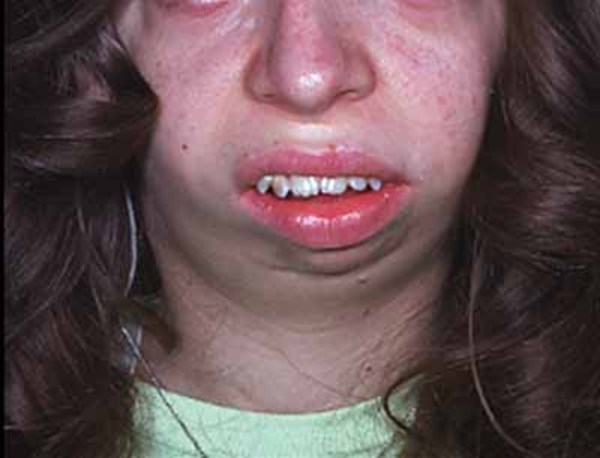
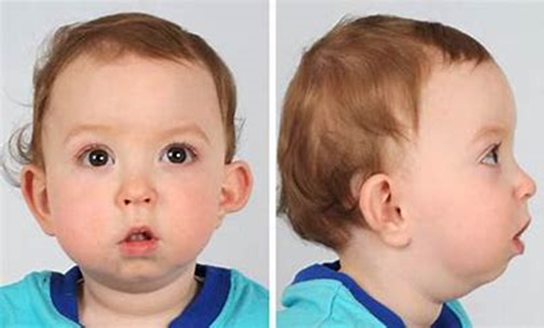
Cherubism, though rare, is a genetic disorder that can significantly affect a person’s quality of life. It is primarily characterized by abnormal bone remodeling in the jaw area, leading to facial swelling and distinct physical features. This rare condition, named for the cherubic appearance it gives affected individuals, is more than just a cosmetic concern—it is a serious disorder with potentially lifelong health and psychological impacts.
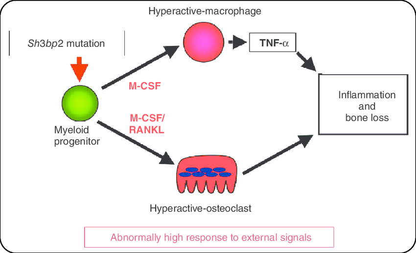
Cherubism is classified as an autosomal dominant disorder, meaning that it can be passed from parent to child with just one copy of the mutated gene. Mutations in the SH3BP2 gene, which plays a role in regulating inflammation and bone homeostasis, are typically responsible for this condition. While cherubism usually manifests in early childhood, its symptoms can vary in severity and progression.
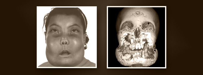
Cherubism is marked by the overgrowth of fibrous tissue in the jawbones, particularly the mandible and maxilla. This tissue gradually replaces normal bone, leading to cyst like spaces within the jaw. This abnormal bone remodeling causes swelling, which may worsen during childhood but can stabilize or even regress as the patient reaches adulthood.
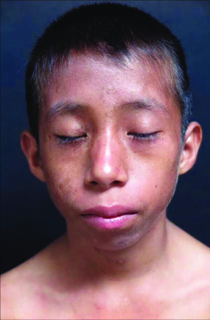
The physical changes that occur with cherubism are profound. Bilateral jaw enlargement results in characteristic facial fullness, often giving children a cherubic appearance. However, this can cause more than just cosmetic concerns. In some cases, the swelling can affect dental development, leading to displaced or absent teeth, and in more severe forms, even interfere with speech, chewing, and breathing.
Despite the seemingly benign facial changes associated with cherubism, the condition can lead to several complications:
As cherubism is a genetic condition, current treatments focus on managing the symptoms rather than curing the disorder. In milder cases, the disease may stabilize on its own with no need for treatment. For more severe cases, interventions may include:
Recent research efforts have delved deeper into the molecular pathways that contribute to cherubism, with the hope of identifying more targeted therapies. Understanding the role of the SH3BP2 gene and its associated pathways may offer potential for future treatments aimed at preventing or reversing the bone changes seen in cherubism.
At our organization, we are committed to advancing the understanding of cherubism through cutting edge research. Our goal is to unravel the complexities of this rare disorder and explore innovative approaches for therapeutic intervention. As we expand our knowledge of gene based disorders, we aim to improve the quality of life for those affected by cherubism and similar conditions.
Cherubism is not just a cosmetic anomaly—it’s a serious condition that requires a comprehensive approach to treatment and care. By raising awareness and supporting ongoing research, we hope to provide better solutions for individuals living with cherubism and other rare diseases.
Research Assistant (exRNA Therapeutics)
The human immune system is a complex defense network that protects the body from infections and diseases. However, in some rare cases, genetic mutations can impair this defense system, leading to severe immunodeficiencies. One such condition is ZAP70 deficiency, a form of Severe Combined Immunodeficiency (SCID), which hinders the immune system's ability to combat infections. ZAP70 deficiency is an extremely rare, yet critical, genetic disorder that profoundly impacts the adaptive immune response, leaving individuals vulnerable to life-threatening infections.
ZAP70 deficiency is a genetic disorder caused by mutations in the ZAP70 gene, which encodes the Zeta-chain-associated protein kinase 70 (ZAP-70). This protein plays a crucial role in T cell receptor (TCR) signaling, a pathway essential for the proper activation and function of T cells, a type of white blood cell that fights infections. When ZAP70 is dysfunctional or absent due to mutations in the gene, T cells cannot adequately respond to pathogens, leaving the individual susceptible to infections.
Patients with ZAP70 deficiency often exhibit symptoms early in life, such as chronic infections, failure to thrive, and recurrent fevers. Laboratory tests typically show a lack of T cell activity, despite normal numbers of T cells, as well as low or absent CD8+ cytotoxic T cells, which are vital for directly attacking infected cells. Without treatment, these immune dysfunctions lead to fatal infections
Zeta-chain-associated protein kinase-70 (ZAP-70) is a tyrosine kinase mainly expressed in T cells and NK cells. The ZAP70 gene is located on chromosome 2q12, and it encodes a kinase that is activated following the engagement of the TCR complex with an antigen. The activation of ZAP-70 triggers a cascade of intracellular signaling events, leading to T cell activation, proliferation, and differentiation. This process is critical for mounting an effective immune response.
Mutations in the ZAP70 gene can be missense mutations, nonsense mutations, or frameshift mutations that lead to a non-functional or absent ZAP-70 protein. These mutations impair the T cell signaling pathways, making it impossible for the immune system to effectively identify and fight pathogens.
Treatment for ZAP70 deficiency traditionally involves hematopoietic stem cell transplantation (HSCT), which can restore immune function by providing the patient with healthy, functional immune cells. However, this treatment is not without risks, including graft-versus-host disease, complications from chemotherapy, and limited donor availability.
The search for less invasive and more targeted therapies has led researchers to explore antisense oligonucleotide (ASO) technology, a novel therapeutic approach that could revolutionize the treatment of genetic disorders like ZAP70 deficiency.
Antisense oligonucleotide technology involves the use of short, synthetic strands of DNA or RNA designed to bind to a specific messenger RNA (mRNA) sequence. By binding to the defective mRNA produced by the mutated gene, ASOs can prevent the production of a dysfunctional protein or modulate the expression of a gene. In the case of ZAP70 deficiency, ASO therapy could potentially target the faulty ZAP70 mRNA and correct or mitigate the effect of harmful mutations.
While ASO therapy is still in its experimental stages for many genetic diseases, its target specificity and precision offer significant advantages over traditional treatments. With advancements in gene-targeted therapies, ASOs hold promise as a treatment for ZAP70 deficiency, offering a less invasive and potentially more effective option for patients with this rare immunodeficiency. Clinical trials exploring the efficacy of ASO treatments for other genetic diseases, such as spinal muscular atrophy (SMA), have already shown tremendous success, paving the way for similar approaches in SCID conditions.
ZAP70 deficiency, while rare, represents a critical challenge for those affected by it. As we continue to advance our understanding of genetic diseases and develop cutting-edge therapies, antisense oligonucleotides offer an exciting new avenue for treating ZAP70 deficiency. By targeting the very genes responsible for the condition, ASO therapy could provide a precise and effective treatment, transforming the outlook for patients with this debilitating immunodeficiency.
With ongoing research and the development of personalized medicine, the future looks promising for individuals suffering from ZAP70 deficiency and other genetic diseases, ushering in a new era of gene-targeted therapeutics that could one day offer cures for rare diseases.
Research Assistant (exRNA Therapeutics)
17q11 microdeletion syndrome is a rare genetic disorder which is caused by deletion of Specific regions of Chromosome 17(17q11.2) including the Neurofibromatosis type 1 (NF1) gene region.The NF1 gene encodes the protein neurofibromin,which is involved in several signaling pathways specifically Ras pathway and regulate cell growth and division.NF1 primarilly acts as a tumor suppressor by inhibiting the Ras protein which promotes cell growth and division,which can otherwise lead to tumor formation. It also plays a key role in the differentiation and development of cells, particularly in the nervous system, including nerve and Schwann cells. The 17q12 recurrent deletion is found in 0.002% to 0.007% of people from large groups not chosen based on disease.

Neurofibromatosis type 1 (NF1) is characterized by a diverse range of symptoms affecting multiple body systems. Common symptoms include café au lait spots (flat, light brown patches on the skin, and ). Individuals often develop neurofibromas (benign tumors that can appear on or under the skin with plexiform neurofibromas potentially causing significant swelling).Lisch nodules which are small harmless growths on the iris, are common. Optic pathway gliomas can also form along the optic nerve, possibly affecting vision. Bone issues like scoliosis and leg bowing may arise, along with learning challenges such as ADHD and specific difficulties. There's also a higher risk of heart problems like high blood pressure, as well as neurological symptoms like headaches and seizures
17q11 microdeletion syndrome requires a multifaceted therapeutic approach to manage its symptoms and improve the quality of life for affected individuals.Emerging therapies, such as targeting the RAS/MAPK signaling pathway with treatments like MEK inhibitors, which may help reduce the effects of neurofibromin loss.Current research into gene therapy for 17q11 microdeletion syndrome and neurofibromatosis type 1 (NF1) is exploring several innovative strategies. One key area involves gene replacement therapy, which aims to deliver functional copies of the NF1 gene via viral vectors to restore neurofibromin production. Additionally, researchers are exploring CRISPR/Cas9 gene editing to correct mutations or introduce functional genes directly into affected cells.
Research Assistant (exRNA Therapeutics)
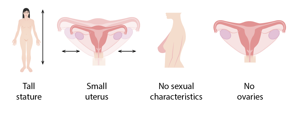
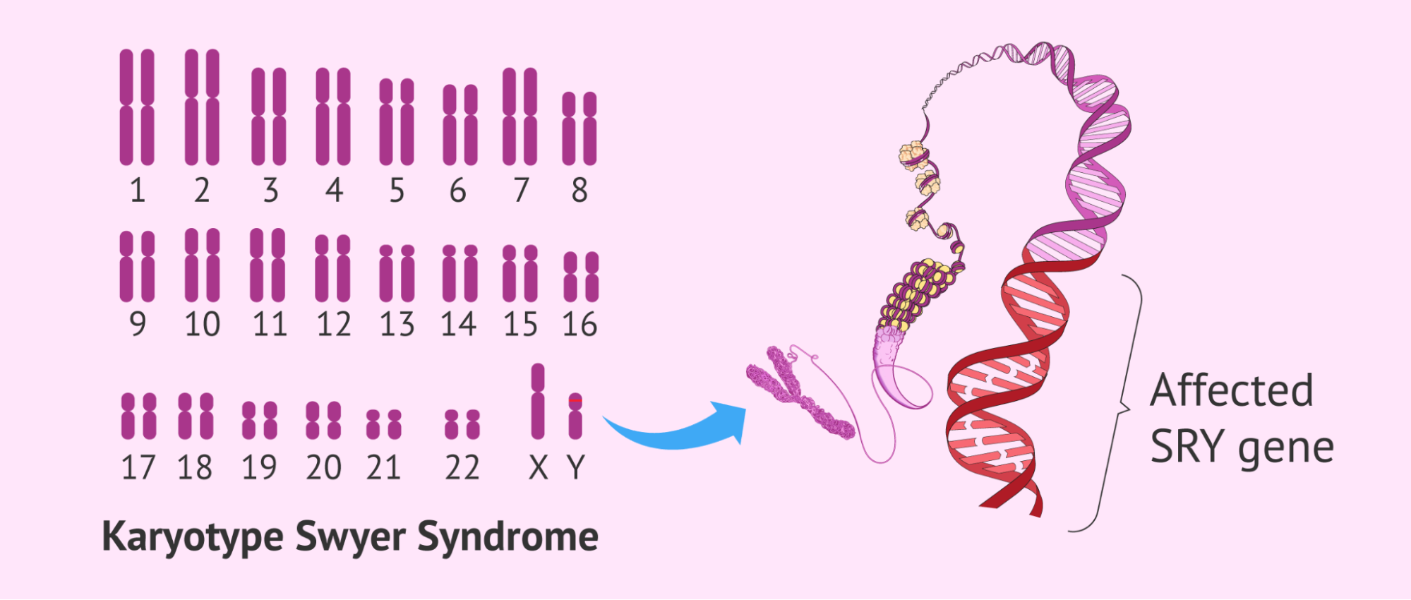
Swyer syndrome, also known as 46 XY complete gonadal dysgenesis is a rare genetic condition that affects sexual development. Individuals having this condition have an XY chromosome pattern and are associated with males. Their body develops as a female's body due to the inability of gonads (testes) to form or function properly.

Gonadal Dysgenesis - It is a condition in which gonads are non-functional and underdeveloped, that is streak gonads which do not produce hormones.
External Female Characteristics - Individuals having this condition typically appear as a female at birth and have normal female external genitalia and secondary sexual characteristics. Individuals do not undergo normal pubertal development due to the absence of ovaries that leads to the lack of estrogen production.
Infertility - Individuals with this condition usually are infertile because their gonads do not function properly.
Risk of Gonadal tumors - It increases the risk of developing gonadal tumors that leads to the surgical removal of dysgenetic gonads.
Causes- - This disorder is primarily associated with mutations in various genes that play critical roles in sexual development. Some key genes are SRY, DHH, NR5A1, MAP3K1, WNT4, CBX2, GATA4, DMRT1 and so on.
It can be diagnosed by many ways like-
1. Karyotyping
2.Hormonal assessments
3. Pelvic ultrasound or MRI

Hormonal Replacement Therapy- Hormone Replacement Therapy is a crucial component in the management of Swyer syndrome. It involves the administration of estrogen and progesterone to induce puberty and development of secondary sexual characteristics like breast development and also maintain bone health by preventing osteoporosis.
Surgical Gonadectomy- In this condition, surgical removal of gonads is often recommended due to the risk of developing gonadal tumors from streak gonads.
Psychological Support- Psychological support is an essential aspect of care for individuals with Swyer syndrome, as it provides emotional support and information regarding the genetic implications of the condition. This can help families understand the nature of the syndrome and make informed decisions regarding treatment options.
Research Assistant (exRNA Therapeutics)
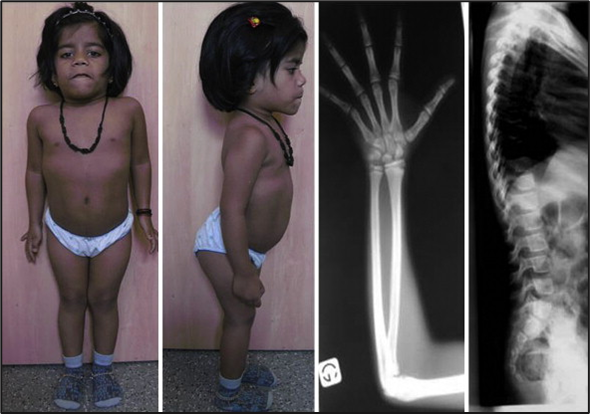
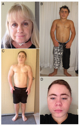
3M Syndrome is a rare genetic condition that causes short stature and specific facial and skeletal features. Although uncommon, it's important to understand this disorder for those affected and their families.
3M syndrome gets its name from the three researchers—Miller, McKusick, and Malvaux—who first described it. This genetic disorder primarily affects growth, leading to significantly shorter height than average, but does not affect intelligence or cognitive development.
One of the key things to know about 3M syndrome is that it’s a congenital condition, meaning it’s present from birth. While some physical features are noticeable early on, other traits develop over time, making early diagnosis and medical management crucial.

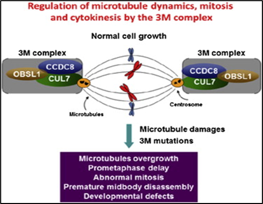
3M syndrome is caused by mutations in three specific genes: CUL7, OBSL1, and CCDC8. These genes are essential in regulating cell growth and development, particularly in skeletal and bone formation.
The condition is inherited in an autosomal recessive pattern. This means both parents must carry a copy of the defective gene, even though they may not show any symptoms. When both parents pass down the mutated gene, the disorder affects the child.

Individuals with 3M syndrome typically exhibit several key symptoms:
A diagnosis of 3M syndrome usually begins with a physical examination to observe growth patterns, skeletal structure, and facial characteristics. However, genetic testing is required to confirm the presence of mutations in the CUL7, OBSL1, or CCDC8 genes. In some instances, X-rays or other imaging techniques may be employed to identify skeletal abnormalities associated with the disorder.
There is currently no specific cure for 3M syndrome. However, the condition can be managed through a combination of medical care and therapies that focus on addressing symptoms rather than altering the underlying genetic causes. Here are the key aspects of treatment:
Individuals with 3M syndrome typically live normal, fulfilling lives despite their physical challenges. They often achieve developmental milestones at the same rate as their peers in terms of intellectual and social skills, although physical mobility and growth may be slower. Families need to assemble a supportive care team, including pediatricians, orthopedic specialists, and therapists, to ensure individuals receive comprehensive care. While short stature and skeletal abnormalities may require long-term medical management, many individuals with 3M syndrome can fully participate in work, education, and social activities.
Our organization is dedicated to advancing the fields of science and medicine, particularly in the realm of rare genetic disorders such as 3M syndrome.. Our mission is to instill hope in individuals and families impacted by rare disorders by exploring innovative therapeutic strategies and leveraging the latest advancements in genetic medicine. Through collaboration with researchers worldwide and ongoing investment in pioneering technologies, we are determined to pave the way for breakthroughs that can significantly improve patient outcomes.
3M syndrome is a lifelong condition. However, ongoing genetic research may offer hope for new treatments in the future. Studies on genetic mutations and their effects on growth may lead to targeted therapies that can help manage or alleviate the effects of the syndrome.
Research Assistant (exRNA Therapeutics)
Understanding disease : Is a subtype of congenital myopathy, a group of muscle diseases present from birth. Multicore myopathies, including CMYO1B, are diagnosed based on the presence of multiple small, damaged regions called “cores” within muscle fibers. These cores disrupt the normal structure and function of muscles, leading to weakness, fatigue, and impaired movement. A key feature of CMYO1B is external ophthalmoplegia—paralysis of the extraocular muscles that control eye movement. This leads to difficulties in maintaining proper eye alignment and impairs visual tracking. Patients also experience generalized muscle weakness, which can range from mild to severe and, in some cases, affect respiratory muscles, increasing the risk of breathing problems.
Genetic Causes: CMYO1B is caused by mutations in specific genes, the most commonly implicated being:
RYR1 as a Therapeutic Target: Given the central role of RYR1 mutations in CMYO1B, targeting this gene provides a focused and logical approach to treatment. Antisense oligonucleotides (ASOs) are an innovative therapy designed to address the genetic defects caused by mutations in genes like RYR1. ASOs are short, synthetic molecules that bind to RNA, modifying the gene expression or splicing process in a way that can correct or mitigate the effects of the faulty gene.
For RYR1-related CMYO1B, ASO therapy offers a unique opportunity to address the root cause of the disease by restoring proper calcium handling within muscle cells. By targeting the specific RYR1 mutations responsible for the disease, ASOs can potentially restore normal muscle function, prevent the formation of abnormal cores, and reduce muscle weakness, ultimately improving quality of life for patients.
The Need for Targeted Therapy in CMYO1B: CMYO1B is a progressive disease that worsens over time, leading to increasing levels of muscle weakness, loss of mobility, and, in severe cases, respiratory difficulties. Given the severity of the condition and the lack of existing treatments, there is an urgent need for therapies that can specifically target the underlying genetic defects.
Challenges in Developing RYR1-Targeted Therapy: While the potential of RYR1-targeted ASO therapy is exciting, several challenges remain in developing an effective treatment for CMYO1B:
One of the primary challenges in ASO therapy is delivering the drug to the affected muscle tissues, particularly the extraocular muscles responsible for eye movement. The current delivery systems must be optimized to ensure that the ASOs reach the target muscles in sufficient quantities to be effective.
ASO therapies typically require repeated administration, as their effects are not permanent. Ensuring that the treatment remains effective over the long term, without causing adverse side effects, is a critical consideration. Developing a treatment regimen that balances efficacy with patient convenience and safety is essential.
CMYO1B is a rare disease, meaning that patient populations are small, making it difficult to conduct large-scale clinical trials. However, advances in clinical trial design for rare diseases, including the use of adaptive trials and patient registries, can help overcome these challenges and accelerate the development of effective treatments.
To date, no treatment has been developed for RYR1-related CMYO1B due to a combination of factors, including the complexity of the disease, the challenge of delivering gene-targeted therapies to muscle tissues, and the small patient population. The high cost of research and development for rare diseases has also been a limiting factor, as pharmaceutical companies often prioritize more common conditions with larger patient bases. However, the success of antisense oligonucleotides in treating other genetic disorders, such as spinal muscular atrophy (SMA), has shown that ASO therapies can be highly effective when targeting specific genetic mutations. These breakthroughs in gene-based therapies provide a clear path forward for developing RYR1-targeted treatments for CMYO1B.
The development of RYR1-targeted ASO therapy represents a promising frontier in the treatment of CMYO1B. With advancements in genetic research, RNA-based therapies, and delivery systems, we are now closer than ever to providing a targeted treatment that can address the root cause of this debilitating disease. Collaboration between researchers, clinicians, and patient advocacy groups will be critical in moving this research forward. By increasing awareness of CMYO1B and its impact, we can help drive the funding necessary for clinical trials and accelerate the path to treatment. Furthermore, personalized medicine approaches, where therapies are tailored to individual genetic mutations, will play a crucial role in developing ASOs that are effective for RYR1-related conditions.
Congenital Multicore Myopathy with External Ophthalmoplegia is a serious and progressive disorder that significantly impacts the lives of those affected. Mutations in the RYR1 gene are the primary cause of this condition, leading to disrupted calcium regulation in muscle cells and subsequent muscle weakness. The development of antisense oligonucleotide therapies specifically targeting RYR1 offers hope for a future where patients with CMYO1B can receive precise, effective treatment. While challenges remain, the progress in RNA-based therapies and advancements in genetic medicine provide a clear path forward. With continued investment, research, and collaboration, we may soon see effective treatments that can improve the quality of life for those living with this rare and challenging condition.
Research Assistant (exRNA Therapeutics)
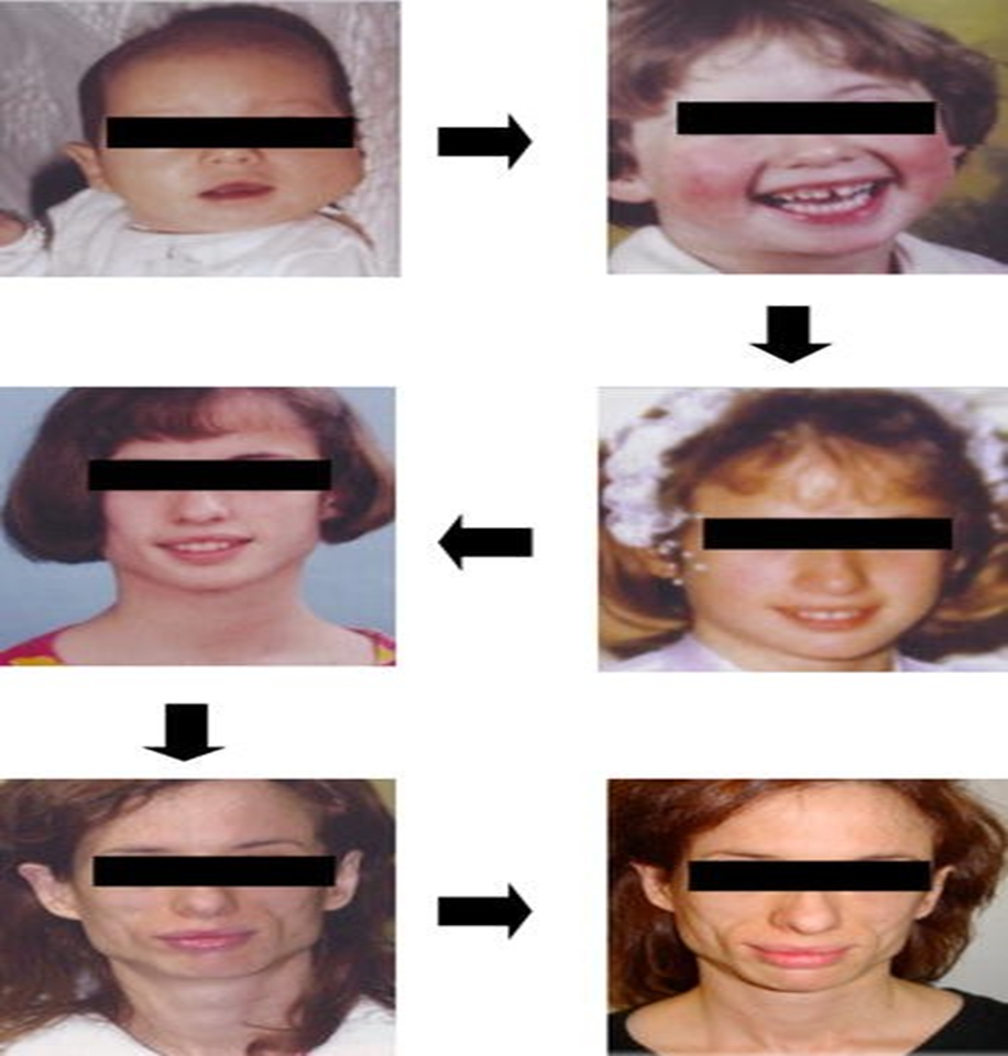
A rare form of Acquired partial Lipodystrophy is also known as Barraquer-Simons Syndrome
Barraquer-Simons syndrome, also known as acquired partial lipodystrophy (APL), is a rare condition characterized by the progressive loss of subcutaneous fat layers, primarily affecting the upper body parts of the body. This blog aims to provide a comprehensive overview of the syndrome, its clinical features, potential causes, associated complications, and treatment options.
Barraquer–Simons syndrome is a form of partial symmetric lipodystrophy, primarily affecting the upper part of the body which is marked by the loss of subcutaneous adipose tissue, particularly in the face, arms, thorax, neck and upper abdomen area while the lower part often retains or even increase their fat storages shown in Figure-1. It is also classified as a type of lipodystrophy, (which refers to a group of disorders marked by abnormal or degenerative conditions affecting adipose tissue). In APL, the loss of fat is typically symmetric and follows an extending from the cranium to the posterior part of the body, starting from the face and neck and potentially extending down to the trunk and thighs. The condition mostly affects women, with a reported ratio of 4:1 compared to men.
The hallmark of Barraquer-Simons syndrome is the gradual loss of subcutaneous fat that can lead to:
Facial Changes: Patients often develop a thin and bony appearance that can be caused by aging, illness, diet, or lifestyle factors due to fat loss in the cheeks and temples (The flat areas that run from the edge of the brows to the tip of the ears and back to the hairline).
Upper Body Fat Loss: This includes the neck, shoulders, and upper arms while sparing lower extremities.
Psychological Impact: The visible changes can lead to significant psychological distress and impact quality life of an individual affected with this syndrome.
Autoimmune Disorders: There is a notable association with autoimmune diseases like systemic lupus erythematosus in 10-20% of cases observed in these diseases.
Membranoproliferative Glomerulonephritis: Approximately 20% of patients may develop this renal complication years after the onset of lipodystrophy due to alterations in complement pathways such as renal diseases C3 nephritic factor, Autoimmune haemolytic anemia, IgG4–positive multiorgan lymphoproliferative syndrome, Neuromyelitis Optica spectrum disorders etc.

Barraquer-Simons syndrome, or acquired partial lipodystrophy, has been associated with genetic factors which mainly responsible for the genes related to this syndrome which are mentioned below;
LMNB2: LMNB2 gene may be involved in the pathology of Barraquer-Simons syndrome where mutation is seen to get affected by these diseases. This gene encodes a protein that is crucial for maintaining the structure of the nuclear envelope, and its dysfunction could affect adipose tissue metabolism and integrity.
C3 Nephritic Factor: The presence of C3 nephritic factor (C3NeF) is commonly observed in patients with this syndrome. This autoantibody can cause dysregulation of the complement system, leading to adipocyte lysis and contributing to fat loss.
LMNA (Lamin A/C): Associated with partial lipodystrophy
PPAR (Peroxisome proliferator-activated receptor): Another gene linked to lipodystrophy.
AKT2: nvolved in metabolic processes related to fat storage.
ZMPSTE24: Also associated with partial lipodystrophy.
AGPAT2: BSCL2: CAV1: PTRF; These genes are linked to congenital total lipodystrophy, which is a different form of the disorder.
Diagnosing Barraquer-Simons syndrome involves a combination of clinical evaluation and laboratory tests. Key indicators include:
Management strategies for Barraquer-Simons syndrome focus on addressing cosmetic concerns and any associated complications. Some effective treatments include:
Barraquer-Simons syndrome is a rare but impactful condition that requires careful management due to its physical and psychological effects observed in diseased conditions. Early recognition and intervention can significantly improve patient outcomes, particularly through cosmetic procedures that restore facial volume. Ongoing research into its pathogenesis and treatment options continues to be essential for better understanding and managing this unique form of Acquired partial lipodystrophy.
Research Assistant, exRNA Therapeutics
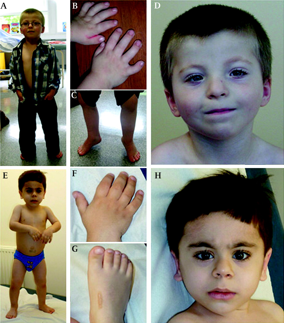
Acromicric dysplasia (ACM) and Geleophysic dysplasia type 2 (GPD2) are rare genetic disorders that share some overlapping features. Both conditions are characterized by severe short stature, short hands and feet, joint limitations, and thickened skin. Both conditions are caused by mutations in the ADAMTSL2 gene, which plays a crucial role in the development and maintenance of connective tissue.Both ACM and GPD2 are extremely rare, with fewer than 30 cases have been reported to date.Their exact prevalence is not well-documented, but they are considered to be part of a broader group of dysplasia syndromes.Acromicric dysplasia is characterized by short stature, short hands and feet, joint stiffness, and distinct facial features like a broad nose , full cheeks, a thin upper lip, and a flat philtrum. Geleophysic dysplasia type 2 shares similar features, including short stature and joint stiffness, but also includes progressive thickening of the skin (skin fibrosis) and heart valve problems. Patients with either disorder often have delayed growth and disproportionately short limbs. Individuals with GPD2 may also have more severe skeletal presentations compared to those with ACM. Additional features can include cardiac valve thickening, tracheal stenosis, bronchopulmonary insufficiency, liver enlargement, and recurrent respiratory and ear infections.Both conditions are caused by mutations in the FBN1 gene, which codes for fibrillin-1, a protein essential for the formation of connective tissue. These mutations disrupt normal bone growth and tissue development. Geleophysic dysplasia type 2 specifically can be caused by mutations in ADAMTSL2 gene, leading to impaired function of the protein it encodes, which is essential for normal connective tissue structure.These mutations are inherited in an autosomal dominant pattern.Treatment: There is no cure for these disorders. Treatment focuses on managing symptoms, such as addressing joint stiffness, skeletal deformities, and heart issues. Physical therapy ,surgical interventions for skeletal deformities, cardiac valve replacement for severe valve thickening, and regular monitoring of cardiovascular health are often recommended to improve quality of life.


Research Assistant, exRNA Therapeutics
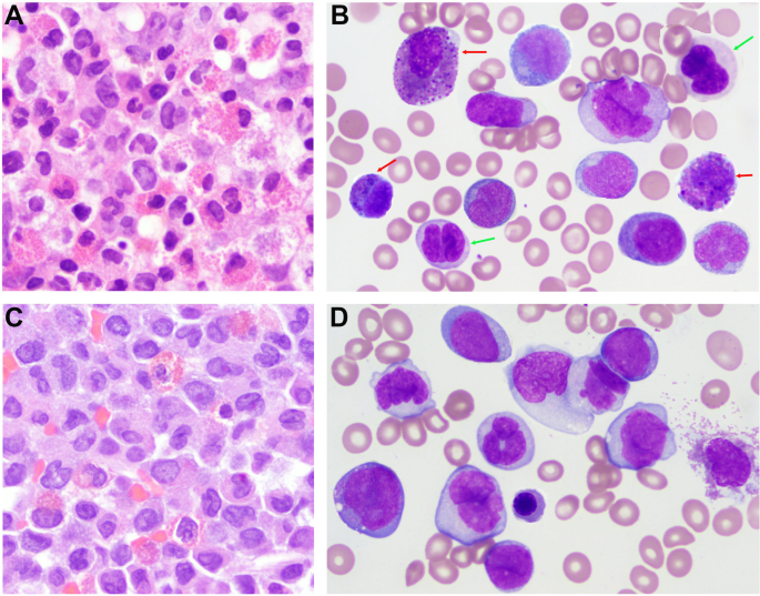
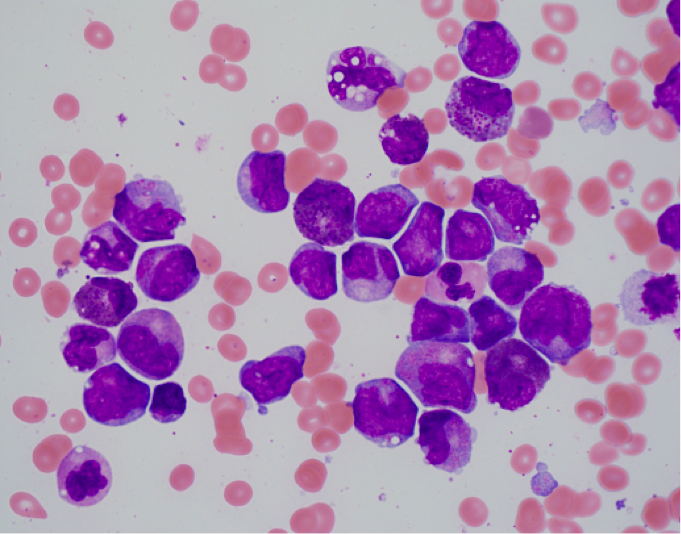
Acute myeloid leukemia (AML) with inversion of chromosome 16 (inv(16)(p13q22)) or translocation t(16;16)(p13;q22) is a subtype of AML classified under the World Health Organization's category of AML with genetic abnormalities.Acute Myeloid Leukemia (AML) with the chromosomal abnormalities inv(16)(p13q22) or t(16;16)(p13;q22), also known as AML with abnormal bone marrow eosinophils, is a rare form of leukemia characterized by the fusion of the CBFB and MYH11 genes. This condition accounts for approximately 5-8% of all AML cases and is typically diagnosed in adults. This variant is more common in younger adults and accounts for about 5-10% of all AML cases. It often presents in patients aged 20-50 years. Patients typically exhibit symptoms associated with bone marrow infiltration, including fatigue, fever, weight loss, recurrent infections due to bone marrow failure, and increased susceptibility to infections due to anemia and leukopenia. Patients may also experience thrombocytopenia, leading to symptoms like pallor and shortness of breath.The presence of eosinophils can also lead to symptoms like skin rashes or respiratory issues.Some patients may present with an enlarged spleen or liver, as well as bone pain.The cause of this condition is the chromosomal rearrangement involving the CBFB and MYH11 genes, resulting from either the inversion (inv(16)) or translocation (t(16;16)). This rearrangement disrupts normal cell development and leads to the uncontrolled growth of abnormal white blood cells.The cause of this subtype involves genetic mutations leading to the abnormal proliferation of immature myeloid cells with eosinophilic differentiation.The inv(16) results in a fusion gene that plays a crucial role in leukemogenesis. Additional mutations such as those in FLT3 or KIT may also be present, influencing prognosis.Treatment typically includes intensive chemotherapy regimens, often followed by consolidation therapy with high-dose cytarabine and anthracyclines. Patients with this chromosomal abnormality tend to respond well to standard AML treatments, and the long-term prognosis is relatively favorable compared to other AML subtypes. Stem cell transplantation may be considered for eligible patients post-remission.


Research Assistant, exRNA Therapeutics
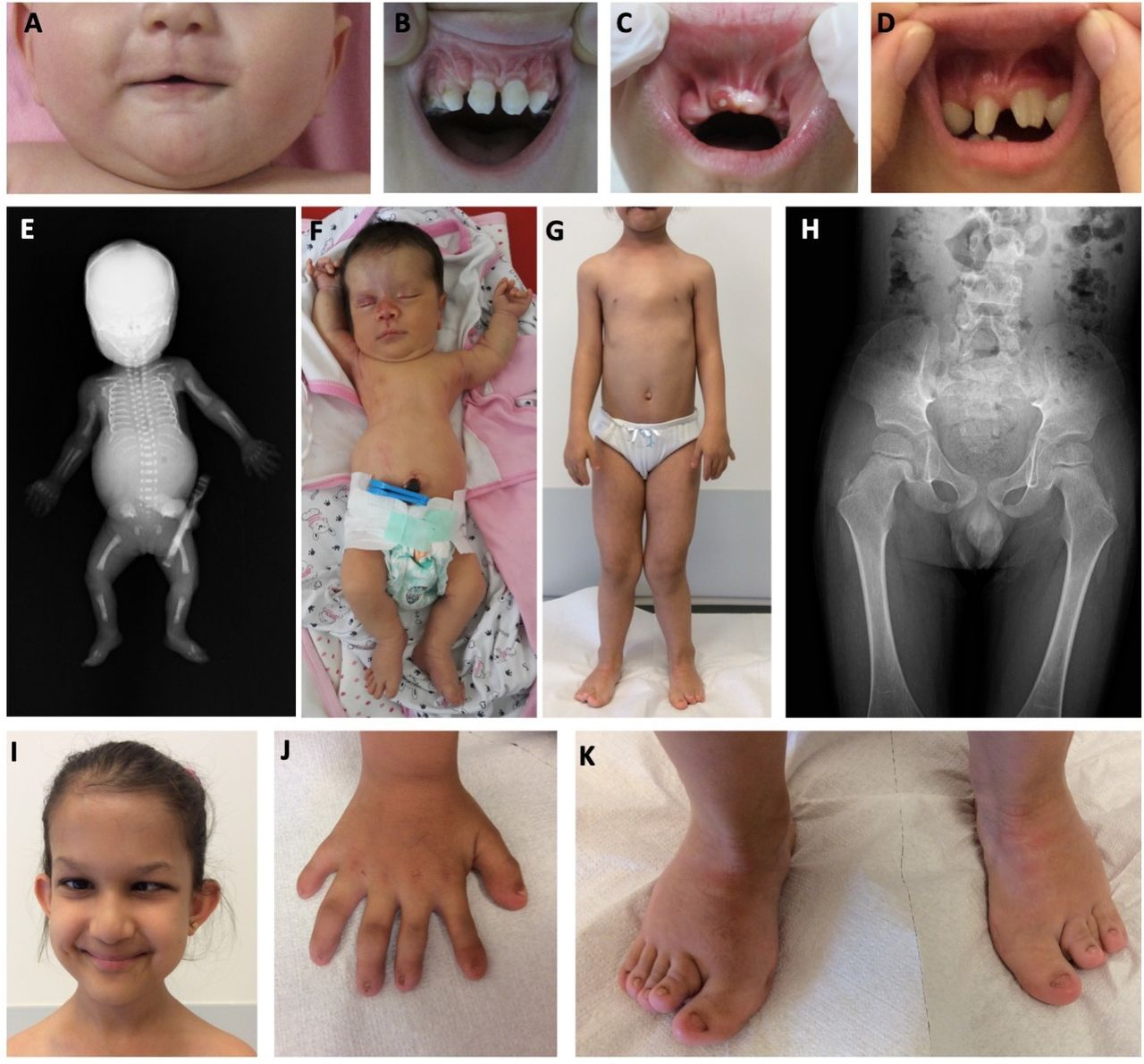
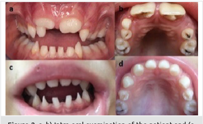
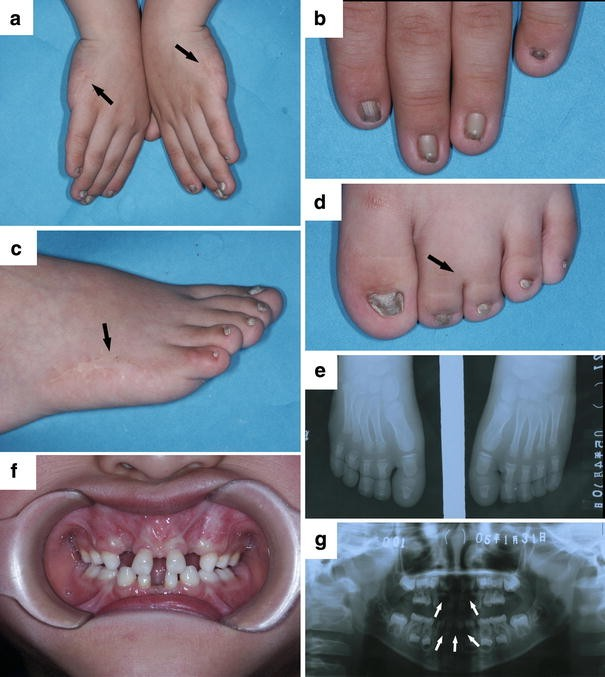
Acrofacial dysostosis, Weyers type (WAD), also known as Weyers acro dental dysostosis or Curry-Hall syndrome, is a rare genetic disorder that affects the development of facial bones, limbs, and teeth. It is part of the acrofacial dysostosis group of conditions, characterized by craniofacial anomalies and limb abnormalities. Prevalence is not well defined, but it is considered a rare disorder with fewer than 50 cases documented globally. It is classified under acrofacial dysostosis syndromes, which are generally uncommon. The main features include craniofacial dysmorphism such as cleft lip and/or palate, absence or hypoplasia of the nose, and ear anomalies. Dental issues are also prominent, with patients often experiencing oligodontia (missing teeth), hypodontia (fewer teeth), peg-shaped teeth, malformations of the lower jaw (micrognathia), and other anomalies affecting the teeth. Skeletal features may include hand and limb malformations. Patients often exhibit onychodystrophy (abnormal nail development), postaxial polydactyly (extra fingers or toes),joint stiffness, heart defects may also be present, and short stature due to moderately restricted growth. Weyers type generally does not affect intelligence.Weyers Acro-Dental Dysostosis is caused by mutations in the EVC and EVC2 genes, which are crucial for the development of bones and teeth. These genes play a role in the Hedgehog signaling pathway. These mutations disrupt the Sonic Hedgehog signaling pathway, critical for the proper formation of bones, teeth, and nails. WAD is inherited in an autosomal dominant manner, meaning only one copy of the mutated gene from an affected parent can lead to the disorder in offspring. It is linked to mutations in the TP63 gene, which is crucial for the development of ectodermal structures. This disorder has no cure, and treatment focuses on managing symptoms. This may include addressing the facial, dental, and skeletal issues. Surgical interventions may be required to correct craniofacial anomalies, orthopedic treatment for limb deformities, and regular monitoring for heart conditions. Dental care is particularly important, often involving orthodontics and restorative dentistry to manage dental anomalies. Genetic counseling is recommended for affected families to understand inheritance patterns and implications for future pregnancies.


Research Assistant, exRNA Therapeutics
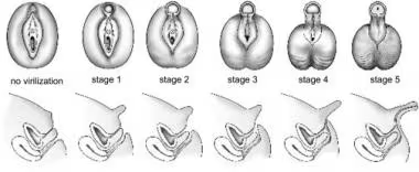
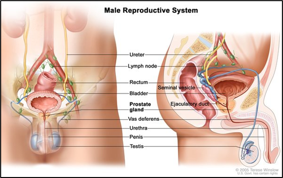
5-alpha-reductase 2 deficiency is a significant cause of 46, XY disorder of sex development (DSD). This deficiency leads to the inability of an individual to convert testosterone into dihydrotestosterone (DHT), a hormone that is crucial for male sexual differentiation. This deficiency arises from mutations in SRD5A2 gene that leads to incomplete masculinization during fetal development and presenting a spectrum of genitals ambiguity at birth.

Role of DHT- Dihydrotestosterone is a hormone essential for normal development of male external genitalia. Individuals having deficiency of 5-alpha-reductase 2 have normal level of testosterone but lack of DHT results in ambiguous genitalia or underdeveloped male genitalia that ranges from a female phenotype with a blind vaginal pouch to a male phenotype having a micropenis and hypospadias.
Clinical Representation-At birth, individuals having this condition are assigned female due to their external genital appearance. During puberty, individuals having this condition experience virilization due to increased testosterone levels that leads to developments in increased muscle mass and changes in voice. Their internal reproductive structures typically remain male, as anti mullerian hormone (AMH), functions normally preventing the development of mullerian structures.
46, XY disorder of sex development due to 5-alpha-reductase 2 deficiency can be diagnosed by combination of clinical assessment and biochemical testing. There are some ways-

Research Assistant, exRNA Therapeutics
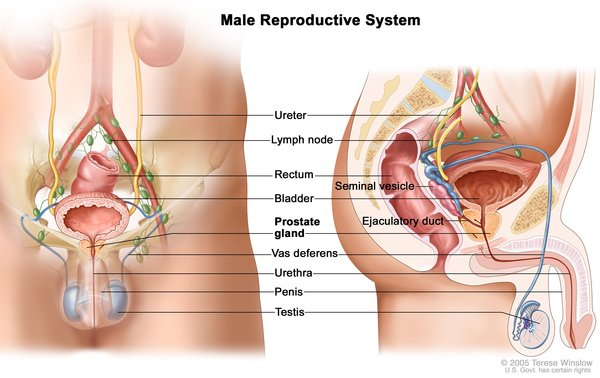
46, XY sex reversal 2 testicular regression syndrome is a rare genetic condition which leads to atypical development of gonads and sexual differentiation in individuals with 46, XY karyotype. This condition falls under the broader category of disorders of sex development (DSDs), where the expected male phenotype does not manifest due to various genetic anomalies.
Testicular Regression Syndrome (TRS) is often considered a part of the clinical spectrum associated with 46, XY sex reversal. This syndrome leads to the failure of testicular development leading to non-functional or absent testes which can result in various degrees of sexual ambiguity. This syndrome emphasizes the importance of early diagnosis and treatment to address potential complications such as infertility and increased risk for gonadal tumors.
Individuals having testicular regression syndrome may present with a range of phenotypes from complete female external genitalia to ambiguous genitalia. Some common features of this syndrome are-
This condition of testicular regression syndrome is linked to mutations in many genes like, AMH, CYP11A1, DMRT1, NR5A1, SOX9, and SRY. These genes are responsible for gonadal development. These genes play a vital role in differentiation of testes and suppression of ovarian development. Disruption in this genetic network leads to incomplete or absent testicular formation resulting in female or ambiguous external genitalia despite the presence of a Y chromosome.
There are many ways to diagnose this syndrome. These are-

There are many ways to treat this syndrome. Some of these are-
Research Assistant, exRNA Therapeutics
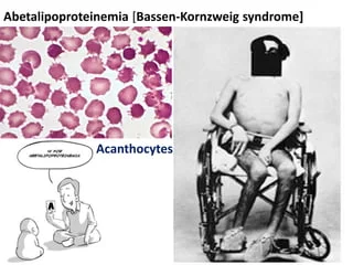
Abetalipoproteinemia is also known as Bassen-Kornzweig Syndrome. It is a rare inherited disorder which results in the body's inability to absorb dietary fats and fat soluble vitamins due to a deficiency in apolipoprotein B.

The condition typically presents in infancy with symptoms such as:
As the individual ages, additional symptoms may develop, including:

It is primarily caused by the mutations in the microsomal triglyceride transfer protein (MTTP) gene that leads to significant malabsorption issues and various clinical manifestations. It is an autosomal recessive pattern. Defects in MTTP gene disrupts the synthesis and secretion of microsomal triglyceride transfer protein (MTP) which are essential for creating beta lipoproteins or apolipoprotein B that transport fats, cholesterol, and fat-soluble vitamins from intestine to bloodstream. Mutations in MTTP can lead to reduced or absent MTP function, causing severely reduced absorption of these nutrients.
Blood Tests- Low or absent levels of lipoproteins like chylomicrons, VLDL, and LDL are characteristics of this syndrome. Acanthocytes in the blood smear are a key sign. Genetic Testing- It confirms mutations in the MTTP gene.
Dietary Management- A diet rich in medium chain triglycerides (MCTs) is absorbed directly into the bloodstream without the use of chylomicron formation, is atypically recommended. Vitamin Supplementation- High doses of fat-soluble vitamins (especially Vitamin E) is crucial to prevent complications related to deficiency. Early diagnosis and treatment can prevent further neurological deterioration and improve long-term outlook.
Research Assistant, exRNA Therapeutics
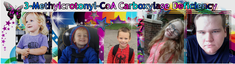
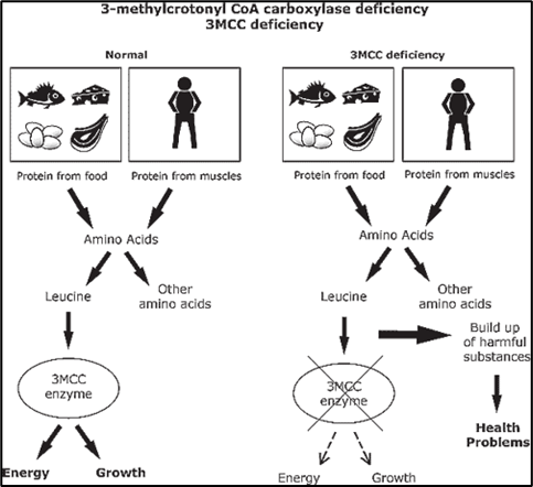
3-Methylcrotonyl-CoA carboxylase deficiency is a rare inherited disorder of amino acid metabolism. It is characterized by the body's inability to properly break down the amino acid leucine due to a deficiency in the enzyme 3-methylcrotonyl-CoA carboxylase. This condition is classified as an organic acid disorder, leading to the accumulation of harmful organic acids and toxins in the body.

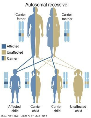
Deficiency of 3MCC is caused by mutations in the MCCC1 or MCCC2 genes, which encode the two subunits of the enzyme 3-methylcrotonyl-CoA carboxylase. This enzyme plays a critical role in the leucine catabolism pathway, breaking down leucine into smaller molecules that the body can use for energy. The condition follows an autosomal recessive inheritance pattern, meaning that a child must inherit two copies of the mutated gene—one from each parent—to manifest the disorder. Carriers (those with one mutated gene) typically do not show symptoms, but they can pass the condition to their offspring. When 3MCC is deficient or absent, toxic intermediates such as 3-methylcrotonylglycine and 3-hydroxyisovaleric acid accumulate in the blood and tissues, leading to metabolic disruption.

3 MCC deficiency is inherited in an autosomal recessive pattern, which means that an affected individual must inherit two copies of the mutated gene, one from each parent. Parents of affected individuals are usually carriers and do not exhibit symptoms of the disorder. However, each child born to two carrier parents has a 25% chance of being affected.

The clinical spectrum of 3MCC deficiency varies widely, with many individuals remaining without symptoms throughout their lives. However, some may experience episodes of illness triggered by metabolic stress, such as fasting, illness, or high-protein diets. These episodes may present with symptoms such as:
- Hypotonia (low muscle tone)
- Vomiting
- Lethargy
- Seizures
- Developmental delays
- Metabolic acidosis (excess acid in the body)
In severe cases, untreated 3MCC deficiency can lead to coma or even death, especially in young children. Therefore, early diagnosis and intervention are crucial for managing the disorder.
Diagnosis usually involves newborn screening programs that detect elevated levels of specific organic acids in urine, such as 3-hydroxyisovaleric acid and 3-methylcrotonylglycine. Confirmatory testing may include measuring enzyme activity in fibroblasts or white blood cells.
Individuals with 3MCC deficiency can lead relatively normal lives with proper management, as there is no cure for the condition. Treatment often involves dietary restrictions to limit the intake of leucine, thereby reducing the accumulation of toxic intermediates. Carnitine supplements are sometimes used to help eliminate excess organic acids from the body.
Individuals with 3MCC deficiency need to avoid fasting and maintain a balanced diet to prevent the occurrence of metabolic crises. In cases of illness or stress, hospitalization and emergency management may be required to stabilize metabolic imbalances.
Newborn screening advances have enabled the early detection and treatment of 3MCC deficiency, leading to significantly improved outcomes for affected individuals. Ongoing research into gene therapy and enzyme replacement therapies may offer new treatment options in the future. Our organization is dedicated to conducting cutting-edge research in the treatment of rare diseases like 3MCC deficiency. By focusing on innovative therapeutic strategies, we aim to contribute to the development of more effective treatments that can enhance the quality of life for individuals affected by these conditions. As with many rare genetic disorders, raising awareness of 3MCC deficiency is crucial for promoting better understanding, diagnosis, and management. With the support of medical advancements and patient advocacy, those living with this condition have hope for a brighter future.
Research Assistant, exRNA Therapeutics
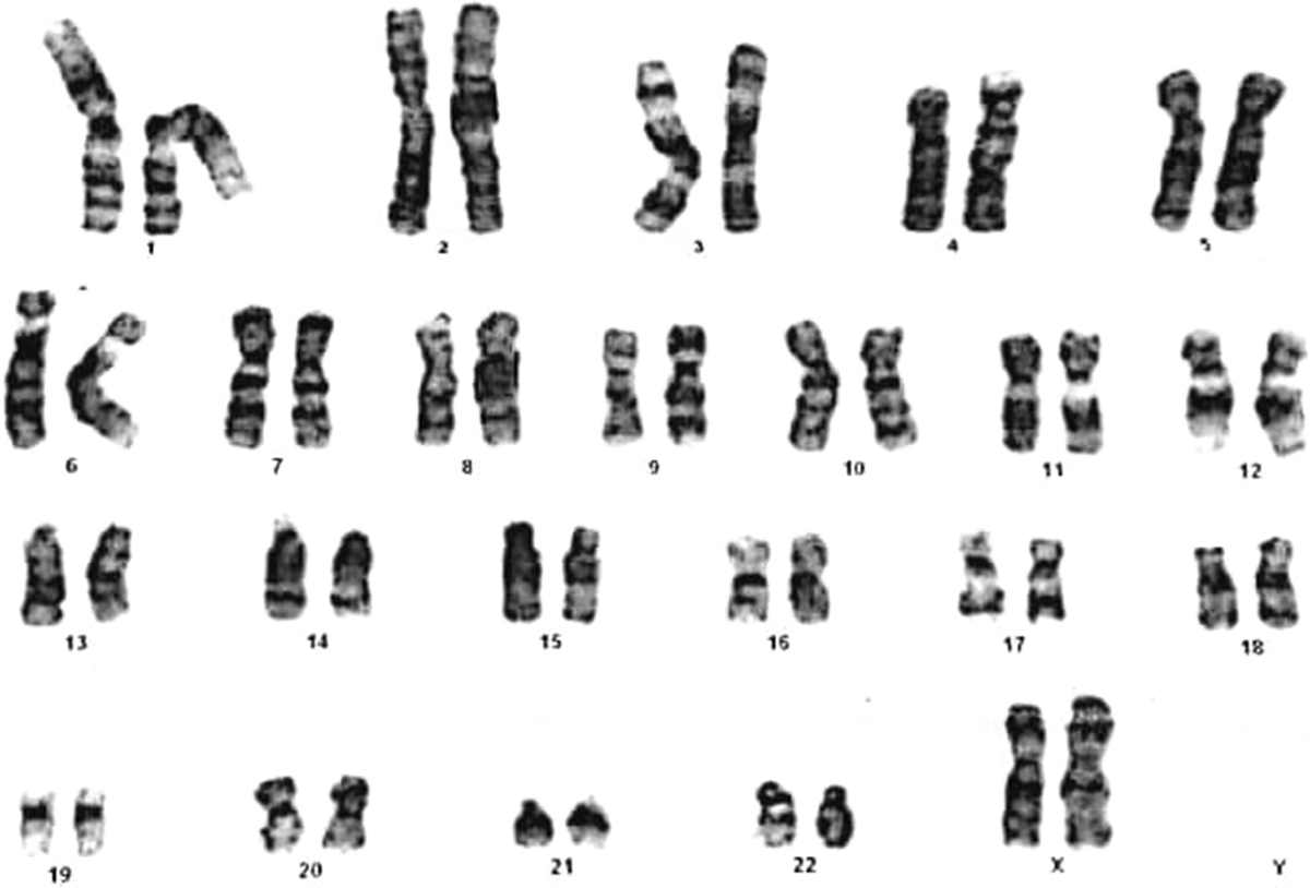
The 46, XX Testicular Disorder of Sex Development (DSD) is a rare condition where individuals with two X chromosomes (typically associated with female development) develop male characteristics. This condition is a form of intersex variation, where there is a discrepancy between chromosomal sex (XX) and phenotypic sex (male). Although individuals affected by 46, XX testicular DSD appear male at birth, they have an atypical genetic makeup.

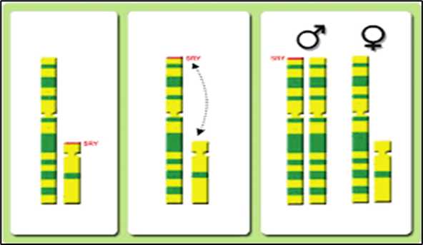
In a typical male, the presence of a Y chromosome triggers the development of male gonads (testes) and the secretion of testosterone, essential for male sexual differentiation. In individuals with 46,XX testicular DSD, the Y chromosome is absent, but male characteristics still develop.The condition is primarily linked to the presence of the SRY gene, which is crucial for testis differentiation. Approximately 80% of cases are SRY-positive, meaning that the SRY gene has been translocated from a Y chromosome segment to an X chromosome during sperm formation. The remaining cases are SRY-negative, where individuals may still develop testicular tissue through unknown mechanisms.

Individuals with 46, XX testicular DSD typically present with:
In rare instances, 46, XX individuals may have ambiguous genitalia or a mix of male and female physical traits.
The condition is often detected in childhood or adolescence, especially if there are concerns about delayed puberty or small testicular size. Sometimes, the diagnosis is made during fertility evaluations, as most affected individuals are infertile.
The diagnosis typically involves:
- Karyotype Analysis: Confirming the 46, XX karyotype.
- Hormonal Assessments: Evaluating levels of testosterone, follicle-stimulating hormone (FSH), and luteinizing hormone (LH) to assess gonadal function.
- Imaging Studies: Ultrasound may be used to evaluate gonadal position and structure.
The management of 46, XX testicular DSD depends on the individual's specific needs, particularly regarding fertility and hormonal issues. Common treatment strategies include:
- Testosterone Replacement Therapy: This is initiated at puberty to promote male secondary sexual characteristics and prevent gynecomastia.
- Psychological Support: Counseling may be beneficial for affected individuals and their families to address identity and social challenges.
- Surgical Interventions: Procedures may be considered for conditions such as hypospadias or gynecomastia when necessary.
Individuals with 46, XX testicular DSD are typically raised as males and identify as male due to their male genitalia and secondary sexual characteristics. However, it's important to recognize that gender identity is a personal matter, and providing psychological support is crucial to help individuals explore and express their gender identity in a supportive environment.
The treatment landscape for 46, XX Testicular Disorder of Sex Development (DSD) is rapidly evolving with the advancement of genetic and personalized medicine. Traditionally managed through hormone therapy and psychosocial support, the future holds the potential for more targeted and individualized treatments. Our organization aims to develop innovative therapies that not only manage but potentially cure 46,XX testicular DSD, improving outcomes and the quality of life for those affected by this rare disorder.
Research Assistant, exRNA Therapeutics
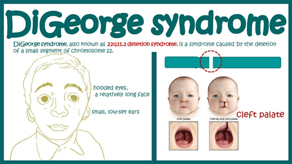
22q11.2 Deletion Syndrome (22q11.2DS) is one of the most common chromosomal disorders, affecting about 1 in 4,000 live births. It is also known by several names, including DiGeorge Syndrome and Velocardiofacial Syndrome (VCFS), reflecting the diverse spectrum of symptoms it causes.
22q11.2 deletion syndrome is a genetic condition caused by the deletion of a small part of chromosome 22. This deletion leads to improper development of several body systems, resulting in a wide range of potential symptoms and complications. The genetic deletion can happen sporadically (without a family history) or be inherited from a parent with the deletion.

The severity of 22q11.2 deletion syndrome varies greatly between individuals. Some of the most common symptoms and complications include:
In most cases, 22q11.2 deletion syndrome is caused by the deletion of a 3 million base pair segment on the long arm of chromosome 22. This deletion occurs randomly during early fetal development in about 90% of cases. The deletion includes the TBX1 gene, which plays a key role in the development of the heart, palate, parathyroid gland, thymus, and facial structures. The wide variability in symptoms is likely due to genetic modifiers and other factors.

Diagnosis is suspected based on clinical symptoms and confirmed by genetic testing techniques like FISH, MLPA, aCGH, or SNP microarrays. Prenatal diagnosis is possible in some cases. Treatment involves a multidisciplinary approach to manage the various symptoms and complications. This may include:
Ongoing research is exploring new ways to manage and treat 22q11.2DS. Gene therapy, advanced diagnostics, and early intervention strategies hold promise for improving the lives of those affected by this condition. With better understanding and awareness, early diagnosis, and comprehensive care, individuals with 22q11.2DS can lead fulfilling lives despite the challenges they may face.
Research Assistant, exRNA Therapeutics
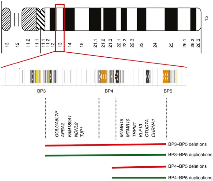
Burnside-Butler syndrome is a rare genetic disorder caused by the deletion of a specific region on chromosome 15, specifically in the proximal long arm region 15q11-q13, which includes five breakpoint sites, most notably BP1 through BP5.This deletion affects four genes which is situated between BP1 to BP5 region: TUBGCP5 (tubulin gamma complex component 50, CYFIP1(cytoplasmic FMR1-interacting protein 1), NIPA1(non-imprinted in Prader–Willi/Angelman syndrome region protein 1), and NIPA2 (non-imprinted in Prader-–illi/Angelman syndrome region protein 2 ). Being an autosomal dominant genetic disorder with incomplete penetrance, exhibits a wide range of symptoms and variability in its expression among affected individuals.The prevalence of the 15q13.3 recurrent deletion in the general population is estimated at 1:5,500.

The key clinical features in Burnside-Butler syndrome includes:
There are no specific drugs for this syndrome. Burnside-Butler syndrome treatments largely emphasize magnesium supplementation.This approach is based on the involvement of the NIPA1 and NIPA2 genes, which encode magnesium transporters and insufficient magnesium levels may lead to issues like seizures and cognitive difficulties. Additionally, psychiatric medications may be prescribed to manage related symptoms, including SSRIs for anxiety and depression, stimulants like methylphenidate for ADHD, and atypical antipsychotics for more severe manifestations. Supportive therapies such as behavioral, speech, and occupational therapy are also essential for addressing developmental delays and behavioral challenges. Overall, while magnesium supplementation appears promising, further research is crucial to establish standardized treatment protocols for this syndrome.
Research Assistant, exRNA Therapeutics
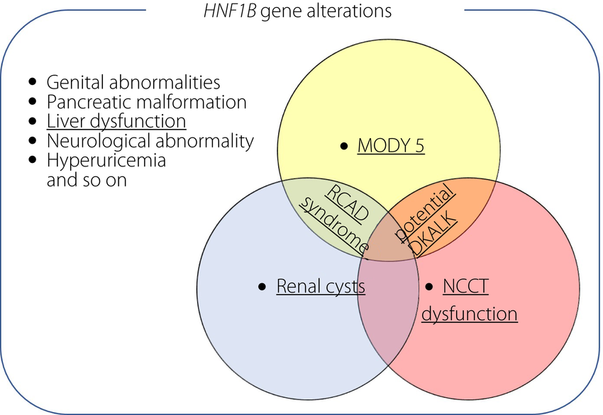
The 17q12 recurrent deletion syndrome is a rare genetic condition caused by the deletion of a specific position(q12) at chromosome 17. This deletion affects one of the two copies of chromosome 17 .Estimated prevalence of this syndrome is 1.6 per 100,000 citizens. RCAD is inherited in an autosomal dominant manner, meaning that only one copy of the mutated gene from an affected parent can cause the disorder in the offspring.This syndrome primarily affects the kidneys, pancreas, and brain. the syndrome can characterized by three main types of health issues; which include Kidney and urinary tract abnormalities (85-90% of cases),Maturity-onset diabetes of the young type 5 (MODY5) (40% of cases),Neurodevelopmental and neuropsychiatric disorders (50% of cases).

Clinical Manifestations
Some patients may also develop hyperuricemic nephropathy which is characterized by high uric acid levels, which can lead to kidney damage.
Genetic testing is a crucial tool for identifying this syndrome,as there is no other diagnostics tool available. The HNF1B score helps for identifying HNF1B mutations by using indicators such as kidney abnormalities and low magnesium levels.Genetic counseling is also recommended for individuals suspected of having 17q12 deletion syndrome to discuss any inheritance pattern.
Currently there is no specific treatment for this deletion syndrome but treatments focusing on managing individual symptoms and complications.Insulin therapy is often necessary to manage blood sugar levels for those having MODY5.Regular monitoring of kidney function is crucial, as structural issues like cystic dysplastic kidneys are common.Some individuals may require surgery for serious kidney dysfunction or complications related to urinary tract issues.
Research Assistant, exRNA Therapeutics
Potocki-Lupski syndrome is a complex genetic disorder which is caused by microduplication of approximately 3.7 megabases on the short arm of chromosome 17, at chromosome 17p11.2 involves the RAI1 gene(retinoic acid inducible 1). Other genes involved in this region are SREBF1, DRG2, LLGL1, SHMT1 and ZFP179 which are responsible for normal development. The estimated prevalence of Potocki-Lupski Syndrome is approximately 1 in 20,000 live births.It is inherited in an autosomal dominant but there are some cases of de novo duplication.

Potocki-Lupski Syndrome (PTLS) is characterized by a range of developmental and physical challenges that significantly impact affected individuals.Infants often experience hypotonia, resulting in low muscle tone and struggle with feeding difficulties due to issues with sucking and swallowing, leading to inadequate weight gain. Developmental delays in motor, social, and cognitive skills are typical in PTLS. Many children encounter significant challenges with speech and language. As they grow, some may exhibit hyperactive behaviors similar to ADHD, along with anxiety and aggression.Furthermore, many display traits associated with autism, especially in their communication skills.Cardiovascular anomalies are observed in roughly 40% of individuals with Potocki-Lupski Syndrome.
Different advance genetic testing procedures are involved for identifying the complex syndrome: Chromosomal Microarray Analysis (CMA) can identify the specific size and location of duplications on chromosome 17p11.2,Multiplex Ligation-Dependent Probe Amplification (MLP) can recognize specific copy number variants,Fluorescence In Situ Hybridization (FISH) to visualize specific chromosome regions,Karyotyping for rule out other chromosomal abnormalities.
At present, there are no specific drugs approved for treating Potocki-Lupski Syndrome (PTLS). Management primarily emphasizes supportive therapies to address the diverse developmental, behavioral, and physical challenges associated with the syndrome.Researchers are exploring compounds that target pathways affected by the duplication of the 17p11.2 region .
Research Assistant, exRNA Therapeutics
Schinzel syndrome is also known by other names as ulnar-mammary syndrome which is a rare inherited disorder characterized by a range of developmental and physical abnormalities observed in individuals suffering from these diseases. This disease was First identified by Dr A. Schinzel in the year 1973 where this syndrome was seen to affect both sex males and females equally, with approximately 50 to 75 cases reported in medical literature.
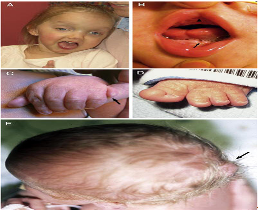
Schinzel-Giedion syndrome is a severe condition that is apparent at birth and affects many body parts and systems. Signs and symptoms of this condition include distinctive facial features, neurological problems, and organ and bone abnormalities it has seen that most affected individuals do not survive past childhood because of their serious health problems
Children with Schinzel-Giedion syndrome can have a variety of distinctive features. In most affected individuals, the middle of the face looks as though it has been drawn inward (midface retraction). Other facial features include a large or bulging forehead; wide-set eyes (ocular hypertelorism); a short, upturned nose; and a wide mouth with a large tongue (macroglossia). Affected individuals can have other distinctive features, including larger than normal gaps between the bones of the skull in infants (fontanelles), a short neck, low-set ears, and an inability to secrete tears (alacrimia).
Schinzel syndrome presents with a variety of symptoms that can significantly impact the quality of life for affected individuals. Some of the most common features include:


Schinzel-Giedion syndrome is caused by mutations in the SETBP1 gene. This gene provides instructions for making a protein that attaches to certain regions of DNA to increase gene activity expression. The SETBP1 protein is found throughout the body, but protein levels are highest during brain development before birth. During this time, nerve cells grow and divide, proliferate and migrate to their proper location in the brain where the SETBP1 protein is thought to control the activity of genes involved in these developmental processes. The SETBP1 gene mutations that have been identified in Schinzel-Giedion syndrome are described as "gain-of-function" mutations because they increase the activity of the SETBP1 protein which increased SETBP1 gene activity likely to alters the expression of other genes that are particularly involved in development before birth.
Diagnosis of Schinzel syndrome is often made through a combination of clinical evaluation and imaging studies. Key diagnostic methods include:
While there is no cure for Schinzel syndrome, management focuses on addressing the individual symptoms and complications. Treatment options may include:
Schinzel syndrome is a complex condition that requires a multidisciplinary approach for management. Early diagnosis and intervention can significantly improve outcomes for affected individuals. Awareness of this rare disorder is essential for healthcare providers to ensure timely support and care for those impacted by its challenges. For families seeking more information or support regarding Schinzel syndrome, connecting with genetic counseling or support groups specializing in rare genetic disorders can provide valuable resources and community support.
Research Assistant, exRNA Therapeutics
Acrodysostosis is a rare congenital disorder characterized by distinct skeletal abnormalities, growth delays, and intellectual disabilities has seen to occur in this disease. This condition encompasses several syndromic presentations, including Maroteaux-Malamut syndrome, which is one of its recognized forms.
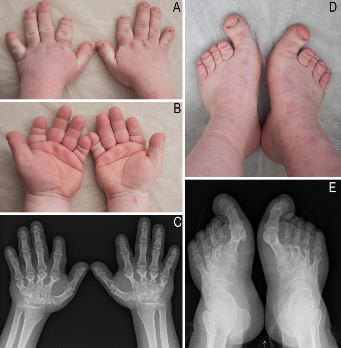
Acrodysostosis primarily affects bone development, leading to features such as observed short stature in Individuals who have significantly shorter arms and legs, also some distinctive facial features which may include a broad, flat nose, wide-spaced eyes, and a protruding jaw bulges due to an improper alignment of the teeth and jaw bones. Some skeletal deformities observed which are characterized by symptoms include short fingers and toes (brachydactyly) and potential deformities in the hands and feet.
The symptoms of acrodysostosis can vary widely among individuals but typically include shows in Figure 1:

Patients affected with acrodysostosis have been found to have genetic defects in one of two distinct genes, those coding for the type 1 regulatory subunit of cAMP-dependent protein kinase alpha (PRKAR1A) or the cAMP-specific phosphodiesterase 4D (PDE4D). Both of these proteins are important components in the GPCR-Gsα-cAMP-protein kinase A (PKA) signaling pathway. In which PKA is the most common effector of cAMP. In the absence of cAMP, PKA exists as a tetramer with two regulatory subunits locking the two catalytic subunits in an inactive state, dominant-negative effect of PRKAR1A or PDE4D mutations for PKA function results in preferential stabilization of mutant regulatory subunits, but preferential release and accelerated degradation of wild-type PRKAR1A and PDE4D subunits, ultimately leading to the suppression of kinase activation leading to these diseases.
Diagnosis usually involves a combination of clinical examination and imaging studies. Key diagnostic indicators include:
Treatment for acrodysostosis is symptomatic and may involve:
Acrodysostosis, particularly its variant Maroteaux-Malamut syndrome, presents significant challenges due to its complex symptoms and genetic basis. Awareness of this condition is crucial for early diagnosis and intervention, which can significantly enhance the quality of life for affected individuals. Support groups and resources are available for families navigating this rare disorder, providing essential information and community support are seen in recent advancements.
Research Assistant, exRNA Therapeutics
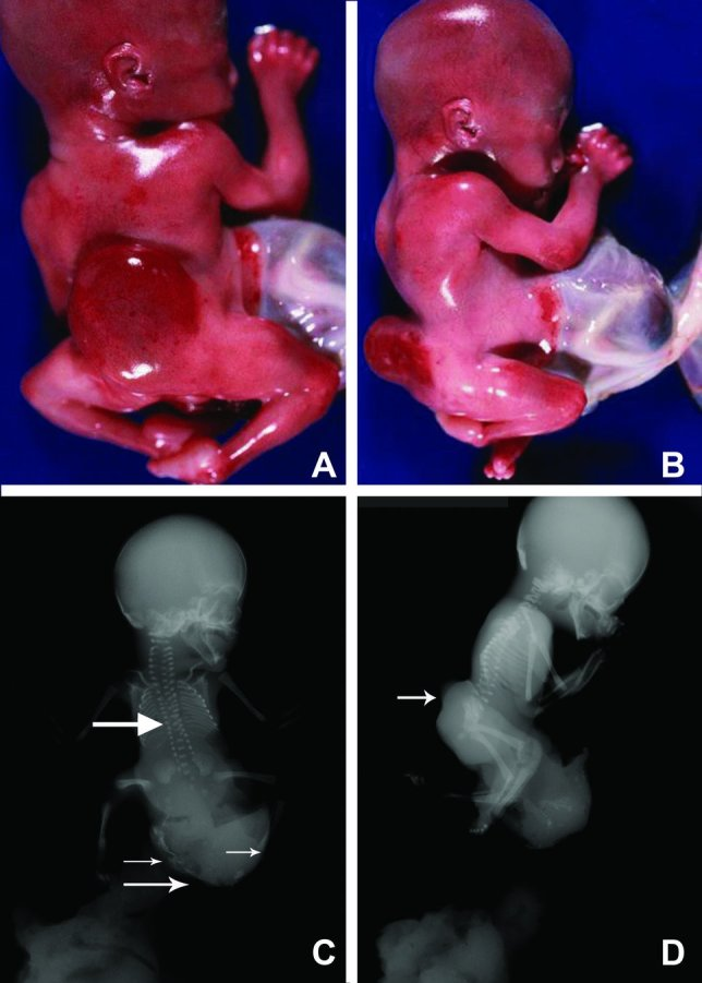
Limb-Body Wall Complex (LBWC) is a rare and severe congenital disorder characterized by a spectrum of debilitating malformations affecting the limbs, thorax, and abdomen area. This condition is often fatal in most of the cases where affected fetuses do not survive to birth or die shortly after delivery.
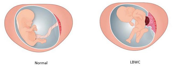
Limb Body Wall Complex (LBWC) is a serious abnormality where it is seen that the baby’s organs are outside of its body and attached to the placenta cell wall. The baby is unable to move or grow properly in that environment and organs are also seen to be damaged. Approximately 250 cases have been reported with this disease so far till date, LBWC is rare and affects approximately 1:15,000 pregnancies.The condition often presents with additional complications such as spinal abnormalities (e.g., scoliosis), renal agenesis, bowel atresia, and various visceral malformations.

The image on the left shows a normal baby and umbilical cord that are freely floating in the amniotic fluid. The baby’s arms and legs are free to move around, and the internal organs are all contained within the body itself. Compared to the image on the right, the baby has LBWC where the baby’s internal organs have come out through a hole from the belly and the organs have stuck to the placental wall and the umbilical cord is very short because of the fixed positioning, as the baby continues to grow it develops abnormal posturing of its spine and limbs in different shape as compared to normal baby as shown in figure-1.
LBWC is marked by multiple congenital anomalies, which may include:

The cause of LBWC have several theories which have been proposed:
LBWC can often be diagnosed prenatally through ultrasound imaging. Key indicators include:
Early detection typically occurs around 11-13 weeks of gestation. In cases where LBWC is suspected, further imaging may be employed to assess the extent of anomalies.
The prognosis for LBWC is difficult due to most affected fetuses not surviving until birth due to the severity of their conditions. For those who survive delivery, immediate medical intervention is often necessary; however even with supportive care survival beyond a few days is rarely seen in the individuals suffering from the diseases. Parents facing a diagnosis of LBWC are often counseled regarding the option of pregnancy termination due to the high likelihood of poor outcomes. If they choose to continue with the pregnancy, close monitoring and additional ultrasounds are recommended to track fetal development and placental health.
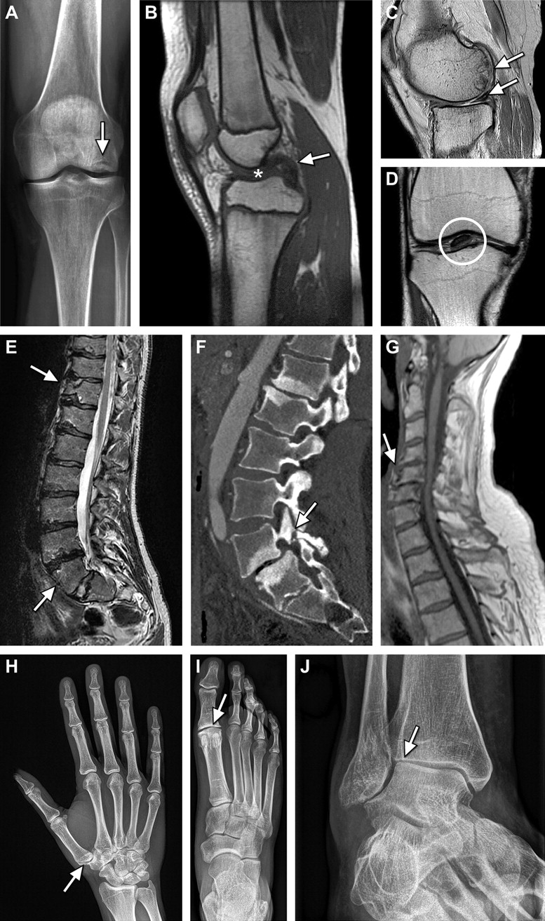
Limb-Body Wall Complex represents one of the most challenging congenital disorders encountered in prenatal medicine. Its complex etiology, severe clinical manifestations, and poor prognosis need a multidisciplinary approach for management and support for affected families. Continued research into its causes and potential interventions remains essential for improving outcomes in future pregnancies.
Research Assistant, exRNA Therapeutics
Aneurysm osteoarthritis syndrome is a rare genetic disorder characterised by the development of arterial aneurysm and early onset arthritis.it is caused by mutation in the SMAD3 gene and is inherited in an autosomal dominant pattern.
Genetic mutations: mutation in the SMAD3 gene disrupts the transforming growth factor beta (TGF-β) Signalling pathway, which plays a crucial role in the development and maintenance of connective tissue.
The SMAD3 gene encodes the SMAD3 protein, a member of the TGF-B path- way that is crucial for TGF-B signal transmission. the effect of SMAD3 mutations on the aortic wall via the histology and

Osteoarthritis in the extremities is characterised by the degradation of articular cartilage and subchondral bone of joints. Osteochondritis dissecans is defined as a separation of an articular cartilage and subchondral bone segment from the remaining articular surface. These abnormalities were already present at a young age and were often the patient's presenting symptom. They seem to be discriminating clinical features in AOS.

A study in 2012 revealed that approximately 40% of AOS patients had long and slender fingers and toes, but overt arachnodactyly was not present Dolichostenomelia was present in 21% of patients. Thorax deformities including pectus carinatum, pectus excavatum, or asymmetry of the costosternal junction, were present in 36% of patients. Scoliosis was present in 61% of our patients, and three of them were operated on for severe scoliosis. One patient had foraminal stenosis requiring foraminotomy L5-S1 With spondylolysis of 14-51. Protrusio acetabuli approximately one-third (35%) of patients and was usually mild. More than 90% of patients had pes planus. Camptodactyly was present in 13% of patients.
Imaging studies: x ray of joints to evaluate the extent of osteoarthritis Genetic testing: confirmation through genetic testing to identify mutations In the SMAD3 gene.
Research Assistant, exRNA Therapeutics
An aortic aneurysm is a bulge that occurs in the wall of the body's main artery, called the aorta. The aorta carries blood from the heart to the body. Aortic aneurysms can occur anywhere in the aorta. They may be tube shaped or round.Aortic aneurysms are relatively common, especially as people get older. They are present in up to 10% of older men and 1–2% of older women.

You're more likely to have an aortic aneurysm if you:
Aneurysms can involve any part of the aorta, but abdominal aortic aneurysms are much more common than thoracic aneurysms. Abdominal aortic aneurysms are 5 to 10 times more frequent in men than in women and have a prevalence of at least 3% in persons older than 50 years.If a descending thoracic aortic aneurysm extends distally and involves the abdominal aorta, a thoracoabdominal aortic aneurysm is created.
Atherosclerosis has long been considered the common underlying cause of abdominal aortic aneurysms. The strength of the aortic wall lies in its extracellular matrix, the most important components of which are elastin and collagen. Consequently, degradation of these structural proteins weakens the aortic wall, allowing aneurysms to develop.
The goal of therapy for patients with aortic aneurysms is to reduce the risk for expansion and rupture of the aneurysm. Although blood pressure control is generally recommended, β-blockers do not reduce the rate of growth of abdominal aortic aneurysms. Size is the major indicator for repair of asymptomatic aortic aneurysms. Abdominal aortic aneurysms larger than 5.5 cm should be repaired,2,3 as should aneurysms larger than 5 cm in good operative candidates. Abdominal aneurysms of 3.5 to 4.4 cm should be monitored every 12 months, and those 4.5 to 5.4 cm should be monitored every 6 months. Descending thoracic aortic aneurysms larger than 6 cm should undergo surgical repair. Aneurysms of the ascending thoracic aorta should be repaired at 5.5 cm .
Abdominal aortic aneurysms less than 4.0 cm have only a 0.3% annual risk for rupture, whereas aneurysms 4.0 to 4.9 cm have a 1.5% annual risk for rupture, and aneurysms 5.0 to 5.9 cm have a 6.5% annual risk for rupture. The overall mortality in patients with abdominal aortic aneurysm rupture is 80%, including 50% mortality even among those who reach the hospital.

Research Assistant, exRNA Therapeutics
Familial thoracic aortic aneurysm and dissection (TAAD) is a genetic condition characterised by the abnormal dilation or weakening of the thoracic aorta, which can lead to life-threatening dissections or ruptures. The genetics of familial thoracic aortic conditions involve several key aspects:
1. Hereditary Patterns:

Research Assistant, exRNA Therapeutics
15q13.3 Microdeletion syndrome is a genetic disorder caused by the deletion of a segment on the long arm of chromosome 15 at position q13.3. This deletion affects at least seven genes, including the important CHRNA7 gene, disrupting critical genetic material and classifying it as a microdeletion syndrome.
Burnside-Butler Syndrome can be passed down through generations in an autosomal dominant pattern or emerge as a new (de novo) genetic anomaly. In the autosomal dominant framework, a single copy of the mutated gene inherited from either parent is sufficient to cause the disorder. Consequently, affected individuals have a 50% chance of transmitting the mutation to their offspring.
The hallmark of 15q13.3 microdeletion syndrome is its broad spectrum of symptoms, which can differ significantly even among family members. Commonly observed features include:
Individuals may experience mild to moderate intellectual disabilities and learning delays.
Social and communication difficulties are prevalent.
Epilepsy, characterized by recurrent seizures, is a frequent concern.
Mental health issues, such as schizophrenia or bipolar disorder, may manifest.
While physical anomalies may exist, no consistent phenotypic pattern defines the syndrome.

The genetic basis of Burnside-Butler Syndrome, or 15q13.3 microdeletion syndrome, is characterized by the deletion of a critical segment on chromosome 15, which involves several key genes, notably NIPA1, NIPA2, CYFIP1, and TUBGCP5. These genes are integral to various aspects of neural development and function.
NIPA1 and NIPA2 are implicated in osmotic regulation and have been shown to influence neuronal growth and differentiation. Their roles in maintaining cellular homeostasis may be crucial for proper brain maturation.
CYFIP1 is associated with synaptic function and plasticity, suggesting its involvement in cognitive processes and behaviors. Alterations in this gene can disrupt neural connectivity, potentially leading to the mental and developmental challenges observed in affected individuals.
TUBGCP5 plays a significant role in microtubule organization, which is essential for cellular structure and function. Disruptions in microtubule dynamics can affect neuronal stability and signaling, further contributing to the neurological manifestations of the syndrome.
Given its varied clinical picture, diagnosing 15q13.3 microdeletion syndrome necessitates a comprehensive approach. Detailed medical histories, thorough physical examinations, and advanced genetic testing techniques, such as chromosomal microarray analysis (CMA), play crucial roles in identifying the condition.
While there is no definitive cure for Burnside-Butler Syndrome, treatment focuses on alleviating symptoms and enhancing the quality of life for those affected:
Customized educational plans can address learning disabilities.
Physical, occupational, and speech therapies are essential for managing developmental delays.
Targeted treatments for epilepsy and mental health conditions are critical.
Patient organizations offer invaluable resources, fostering community support and advocacy.
15q13.3 microdeletion syndrome, or Burnside-Butler Syndrome, affects approximately 1 in 40,000 individuals in the general population. However, its prevalence is notably higher among those with intellectual disabilities or epilepsy, highlighting its significant impact within these communities.
Burnside-Butler Syndrome (15q13.3 microdeletion syndrome) epitomizes the complexity of genetic disorders, with its diverse presentations and unique challenges. By understanding its genetic roots, recognizing its clinical manifestations, and adopting tailored management strategies, we can better support affected individuals and their families. As research progresses, we move closer to unlocking further insights, paving the way for enhanced diagnostic and therapeutic options. Navigating the journey with a rare genetic disorder demands resilience and support. With the right resources and dedicated community backing, individuals with 15q13.3 microdeletion syndrome can lead rich and fulfilling lives.
Research Assistant, exRNA Therapeutics
15q24 microdeletion syndrome, or Chromosome 15q24 deletion syndrome, is a rare genetic disorder caused by deleting a small segment of DNA on the long arm of chromosome 15, typically ranging from 1.7 to 6.1 megabases. The condition affects approximately 1 in 42,000 individuals and results in various developmental challenges and distinct physical features. Most affected individuals share a critical 1.2 Mb region deletion, which is key to understanding the syndrome's genetic implications. The variability in symptoms among individuals highlights the complexity of genetic interactions and their impact on development.
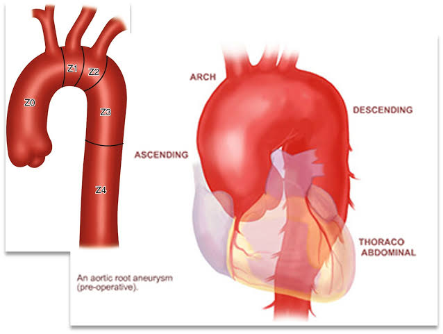
Individuals with 15q24 microdeletion syndrome display a range of symptoms, including:
The 15q24 microdeletion involves deletions ranging from 1.7 to 6.1 megabases on the long arm of chromosome 15, with most patients sharing a critical deleted region of about 1.2 Mb that contains essential genes for normal growth and development.
This deletion causes haploinsufficiency, impairing cellular functions and resulting in various phenotypic effects. The deletion typically arises from nonallelic homologous recombination during reproductive cell formation or early embryonic development, and most cases are de novo mutations, meaning they occur spontaneously rather than being inherited from parents. While the specific genes involved remain largely unidentified, they are believed to play crucial roles in developmental pathways.
Diagnosis of 15q24 microdeletion syndrome typically requires advanced genetic testing methods, such as array comparative genomic hybridization (aCGH) or chromosomal microarray analysis, as conventional karyotyping may miss these small deletions. Most cases are de novo mutations, although some familial cases have been documented. Early and accurate diagnosis is essential for implementing tailored interventions to manage symptoms and support development.
Management of the condition involves an interdisciplinary approach, incorporating specialists from genetics, pediatrics, neurology, speech therapy, and occupational therapy to address the diverse needs of affected individuals. Interventions focus on supporting developmental delays and managing associated medical issues.
15q24 microdeletion syndrome presents various challenges related to growth, development, and health, but early intervention and supportive care can greatly improve the quality of life for affected individuals. Although there is no cure, ongoing research aims to uncover the genetic mechanisms and potential therapeutic targets associated with the syndrome. Families can benefit from support networks that provide information and community connection. The syndrome highlights the complexity of human genetics and emphasizes the importance of increased awareness and collaborative efforts among healthcare providers, researchers, and families to enhance understanding and support for those affected. Through these initiatives, we can foster a more inclusive society that accommodates individuals with genetic differences.
Research Assistant, exRNA Therapeutics
Potocki-Lupski syndrome (PTLS), also known as 17p11.2 microduplication syndrome, is a rare genetic disorder caused by a duplication of a segment on the short arm of chromosome 17 (17p11.2). First identified in 2010, it affects about 1 in 25,000 individuals worldwide. PTLS is characterized by a range of developmental, cognitive, and physical symptoms due to the extra genetic material, distinguishing it from Smith-Magenis syndrome, which involves a deletion of the same segment.
Potocki-Lupski syndrome (PTLS) is characterized by a variety of symptoms impacting development and health.
The primary genetic cause of Potocki-Lupski syndrome (PTLS) is a duplication in the 17p11.2 region, which includes the RAI1 gene. This gene is essential for regulating various biological processes, including circadian rhythms, and its extra copy may contribute to sleep disturbances seen in affected individuals. the candidate genes have been identified within the duplicated section, including SREBF1, DRG2, LLGL1, SHMT1, and ZFP179. The duplication typically spans about 3.7 million base pairs but can vary in size, potentially involving other genes that influence the disorder's variability. PTLS is usually inherited in an autosomal dominant pattern, meaning one mutated gene from an affected parent can cause the condition in their offspring. However, many cases occur spontaneously (de novo), without a family history.
Diagnosis of 15q24 microdeletion syndrome typically requires advanced genetic testing methods, such as array comparative genomic hybridization (aCGH) or chromosomal microarray analysis, as conventional karyotyping may miss these small deletions. Most cases are de novo mutations, although some familial cases have been documented. Early and accurate diagnosis is essential for implementing tailored interventions to manage symptoms and support development.
Potocki-Lupski syndrome (PTLS) is primarily diagnosed through advanced genetic testing methods, such as chromosomal microarray analysis (CMA) and multiplex ligation-dependent probe amplification (MLPA), which can detect microduplications on chromosome 17p11.2.
Management of PTLS (Pitt-Hopkins-like syndrome) focuses on individualized care through personalized therapies, including speech and occupational therapy. A multidisciplinary approach involving healthcare providers, therapists, and educators aims to enhance the quality of life for those affected. Since there is no specific treatment, care plans are tailored to each person’s needs and may include:
Speech and language therapy
Physical therapy
Behavioral interventions
Educational support services
Parental counseling and genetic advice are important for understanding the syndrome and potential recurrence risks in future pregnancies. Raising awareness and understanding is essential, as PTLS symptoms can vary widely, necessitating customized interventions for language and behavioral challenges.
Potocki-Lupski Syndrome (PTLS), caused by the microduplication of chromosome 17p11.2, presents unique challenges and opportunities for understanding genetic disorders. The condition is characterized by a range of symptoms, including developmental delays, behavioral issues, and specific health concerns, highlighting the importance of early intervention and a multidisciplinary approach to care. Tailored therapies can significantly enhance outcomes for individuals with PTLS. Ongoing research is essential for unraveling the complexities of the syndrome and developing effective management strategies. By increasing awareness and fostering supportive communities, we can better meet the needs of those affected and their families. Advances in genetic research offer hope for improved understanding and management of PTLS, ultimately aiming to enhance the quality of life for individuals with the syndrome.
Research Assistant, exRNA Therapeutics
Abdominal aortic aneurysm (AAA) is a multifactorial disease with a strong genetic component. Since first candidate gene studies were published 20 years ago, nearly 100 genetic association studies using single nucleotide polymorphisms (SNPs) in biologically relevant genes have been reported on AAA. The studies investigated SNPs in genes of the extracellular matrix, the cardiovascular system, the immune system, and signalling pathways. SNPs associated with AAA have already been identified in these large multicenter studies. One significant association was of a variant in a gene called CNTN3 which is located on chromosome 3p12.3.

An abdominal aortic aneurysm (AAA) is a swelling (aneurysm) of the aorta – the main blood vessel that leads away from the heart, down through the abdomen to the rest of the body.Large aneurysms are rare, but can be very serious. If a large aneurysm bursts, it causes huge internal bleeding and is usually fatal.The bulging occurs when the wall of the aorta weakens. Although what causes this weakness is unclear, smoking and high blood pressure are thought to increase the risk of an aneurysm.AAAs are most common in men aged over 65. A rupture accounts for more than 1 in 50 of all deaths in this group.
In most cases, an AAA causes no noticeable symptoms. However, if it becomes large, some people may develop a pain or a pulsating feeling in their abdomen (tummy) or persistent back pain. An AAA doesn’t usually pose a serious threat to health, but there’s a risk that a larger aneurysm could burst (rupture). A ruptured aneurysm causes massive internal bleeding, which is usually fatal. Around 8 out of 10 people with a rupture either die before they reach hospital or don’t survive surgery. The most common symptom of a ruptured aortic aneurysm is sudden and severe pain in the abdomen.
Among the 176 patients who had an unruptured aneurysm at the time of the initial ultrasound study, the cumulative incidence of rupture was 6 percent after 5 years and 8 percent after 10 years. However, the risk of rupture over five years was 0 percent for the 130 patients with an aneurysm less than 5 cm in diameter and 25 percent for the 46 patients with an aneurysm 5 cm or more in diameter. All 16 patients who had ruptures had aneurysms that were 5 cm or more in diameter at the time of the rupture.These population-based data challenge the clinical perception that aneurysms typically expand at a rate of 0.4 to 0.5 cm per year. Our data also suggest that for aneurysms less than 5 cm in diameter the risk of rupture is considerably lower than has been reported previously. However, the risk of rupture is substantial for aneurysms 5 cm or more in diameter.
Research Assistant, exRNA Therapeutics
It is a clinical neonatal rare disorder with a complicated pattern of inheritance. Babies are born with the absence of certain layers of skin, most often on the scalp but also on the trunk,and arms and legs. The affected area is typically covered with a thin transparent membrane. The skull and underlying areas may be visible and be abnormally developed.
The exact pathogenesis of ACC is unknown.various theories have been proposed, including the incomplete closure of the neural tube or embryonic fusion lines,intrauterine trauma, vascular compromise from placental insufficiency, ischemic or thrombotic event from foetus( the demise of twin foetus in the late first or early second trimester, genetic mutations. Genetics mutation: Mutation in BMS1 gene, which plays a role in skin morphogenesis and also cases of sporadic and autosomal recessive inheritance have also been reported. Mutations in the ribosomal GTPase BMS1 have been identified as one cause of autosomal dominant aplasia cutis congenita., mutations in EOGT and DOCK6 have been identified as causes for autosomal recessive mutations in DLL4,ARHGAP31, RBPJ, and NOTCH1 have been observed in autosomal dominant forms of the syndrome.In this group of aplasia cutis congenita, the scalp lesions tend to be large.

Aplasia cutis congenita is a rare disorder that may be inherited as an autosomal dominant or autosomal recessive trait .Human traits, including the classic genetic disease,are the product of the interaction of two genes, one received from the father and one from mother.
In most cases, an AAA causes no noticeable symptoms. However, if it becomes large, some people may develop a pain or a pulsating feeling in their abdomen (tummy) or persistent back pain. An AAA doesn’t usually pose a serious threat to health, but there’s a risk that a larger aneurysm could burst (rupture). A ruptured aneurysm causes massive internal bleeding, which is usually fatal. Around 8 out of 10 people with a rupture either die before they reach hospital or don’t survive surgery. The most common symptom of a ruptured aortic aneurysm is sudden and severe pain in the abdomen.
Individuals born with Aplasia cutis congenita lack skin in localised areas of the body usually,but not always scalp.In some cases,the trunk arms and legs may also be affected.The affected area usually replaced with a thin transparent membrane.

Measures to prevent the drying out of the membrane by soothing,blend ointment.Antibiotics should be used only if signs of bacterial infection are present.
Research Assistant, exRNA Therapeutics
Aromatic L-amino acid Decarboxylase Deficiency is a rare genetic disorder characterised by deficient synthesis of Dopamine and serotonin. It prevents in early infancy and causes severe developmental disability and lifelong motor, behavioural and autonomic symptoms including oculogyric crisis ,sleep disorder and mood disturbance.
It is caused by mutations in the DDC gene , which encodes an enzyme called aromatic L- amino Decarboxylase.people born with this condition don't make enough ADDC ,an enzyme that helps our brain and nervous system work properly.This results in a lower level of neurotransmitter,like dopamine symptoms include developmental delays , movement and behaviour problems and issues with involuntary bodily functions.

It typically appear in the first few months of life and can include:
Currently,there are no approved therapies for the disease however supportive treatments are offered to manage symptoms.These may include physiotherapy, occupational therapy,speech and language therapy and enteral feeding. Medication can help manage symptoms,but response varies among individuals. Gene therapy has shown potential in clinical trials.
Molecular genetics - proving heterozygous or homozygous disease causing variants in the DDC gene.CSF examination . Serum examination- Decreased AADC enzyme activity
Individuals with aromatic L-amino Decarboxylase deficiency have complex symptoms, including motor behavioural, cognitive and autonomic findings. Sleep disturbances are present in a majority of affected individuals and mood disturbance irritability and anxiety are also common. Brain MRI is typically either normal or may demonstrate nonspecific abnormalities,such as mild diffuse cerebral atrophy. Seizure are uncommon findings, occurring in fewer than 5% of affected individuals.
Research Assistant, exRNA Therapeutics
Arterial Tortuosity syndrome (ATS) is a rare genetic disorder characterised by abnormal twists and turns of arteries, leading to various cardiovascular complications. The syndrome's genetic cause is linked to mutations in the SLC2A10 gene, affecting collagen and elastin synthesis and is inherited in autosomal recessive manner. It is an extremely rare congenital connective tissue disorder, stenosis or elongation and aneurysm in major and medium size arteries and aorta.

ATS is caused by mutation in SLC2A10 gene which provides instructions for making a protein called GLUT10. The level of GLUT10 appears to be involved in the regulation of a process called the transforming growth factor (TGF-β) Signalling pathway. This pathway is involved in cell growth and division and the process by which cells mature to carry out special functions
This condition is inherited in an autosomal recessive pattern which means both copies of the gene each cell have mutations. The parents of an individual with an autosomal recessive condition each carry one copy of the mutated gene, but they typically do not show signs and symptoms of this condition. The exact incidence of ATS is not well established, with only about 106 patients reported in the literature to date.
Molecular genetics - proving heterozygous or homozygous disease causing variants in the DDC gene.CSF examination . Serum examination- Decreased AADC enzyme activity
1. Ophthalmic Manifestation : A study highlighted various ocular changes associated with ATS , including corneal abnormalities and myopia in both affected patient and carriers.
2. Imaging Techniques: Imaging plays a crucial role in diagnosing ATS, especially in identifying arterial abnormalities.
3. Phenotypic Features: studies have identified phenotypic Features and cardiovascular manifestations in newly diagnosed patients, emphasising the variability of symptoms.
The symptoms of Arterial Tortuosity syndrome can vary significantly among individuals and may not all be present in every case. Early diagnosis and a multidisciplinary approach to management are crucial for addressing the various complications associated with this condition.
Research Assistant, exRNA Therapeutics
Autosomal recessive polycystic kidney disease (ARPKD) is a rare and life-altering genetic condition that primarily affects the kidneys and liver. While many have heard of polycystic kidney disease, ARPKD represents a unique subset of this disorder, distinct from the more common autosomal dominant form (ADPKD). Affecting approximately 1 in 20,000 live births, ARPKD has a profound impact on families and individuals diagnosed with the condition. In this blog, we’ll explore what ARPKD is, its symptoms, causes, and treatment options, while shedding light on the challenges faced by those living with this condition.
ARPKD is an inherited disorder caused by mutations in the PKHD1 gene, which regulates the development and function of the kidneys and liver. The disorder is autosomal recessive, meaning a person must inherit two defective copies of the gene—one from each parent—to develop ARPKD. Carriers (those with only one defective gene) typically do not show symptoms but can pass the gene to their children.
In ARPKD, the kidneys develop fluid-filled cysts that interfere with their normal function, leading to enlarged kidneys and progressive kidney damage. In addition, the liver can be affected by fibrosis (scarring), resulting in complications such as liver dysfunction and portal hypertension.
The symptoms and severity of ARPKD can vary widely among individuals. Some children show signs of the disease before birth, while others may not exhibit symptoms until later in childhood or adolescence. The most common symptoms of ARPKD include:
ARPKD is often diagnosed during prenatal ultrasounds if enlarged kidneys or low amniotic fluid are detected. In other cases, the condition is diagnosed shortly after birth or during early childhood when symptoms such as hypertension or kidney dysfunction become apparent.
There is currently no cure for ARPKD, but treatment focuses on managing the symptoms and preventing complications. The approach to treatment depends on the severity of the disease and the organs affected.
Blood pressure control: Managing high blood pressure is crucial, as it can slow down kidney damage. Medications like ACE inhibitors or angiotensin II receptor blockers are often prescribed.
Diuretics: These medications help reduce fluid buildup and manage kidney function.
Dialysis and kidney transplantation: If kidney failure occurs, dialysis or a kidney transplant may be necessary to sustain life.
Managing portal hypertension: Medications and dietary changes can help control the symptoms of portal hypertension. In severe cases, a liver transplant may be required
Respiratory support: In cases of severe lung involvement, newborns may need respiratory assistance, such as mechanical ventilation.
Nutritional support: Special diets, including low-salt or protein-restricted diets, may be necessary to manage kidney or liver function.
Regular monitoring: Regular visits to a nephrologist (kidney specialist) and hepatologist (liver specialist) are essential for ongoing care.
ARPKD is a rare but serious genetic disorder that affects thousands of families worldwide. While there is no cure, early diagnosis and proper management can help mitigate some of the complications associated with the disease. Advances in medical research offer hope that, in the future, individuals living with ARPKD may have access to more effective treatments and improved outcomes.
Research Assistant, exRNA Therapeutics
Rac1, a small GTPase, plays a significant role in various cellular processes and signaling pathways, including those implicated in muscular dystrophies such as Duchenne muscular dystrophy (DMD). Its involvement in inflammation, oxidative stress, and muscle regeneration makes it a critical target for understanding the pathology and potential therapies for muscular dystrophies.
Rac1 has been linked to inflammatory responses in various muscle diseases, including DMD. Inflammation contributes to the progressive deterioration of muscle function in DMD, making the modulation of Rac1 activity a potentially beneficial strategy in managing this aspect of the disease. By targeting Rac1 signaling pathways, it may be possible to mitigate muscle inflammation and its detrimental effects.
In muscular dystrophies, oxidative stress is a significant contributor to muscle damage and degeneration. Rac1 is known to participate in the production of reactive oxygen species (ROS), which exacerbate oxidative stress. Thus, interventions aimed at inhibiting Rac1 may help reduce oxidative damage in the muscle tissue of individuals with DMD.
Besides its role in promoting inflammation and oxidative stress, Rac1 is also involved in muscle regeneration processes. Effective signaling through Rac1 is crucial for muscle cell fusion and repair mechanisms. Enhancing Rac1 signaling during muscle regeneration could lead to improved therapeutic strategies for patients suffering from muscular dystrophies.
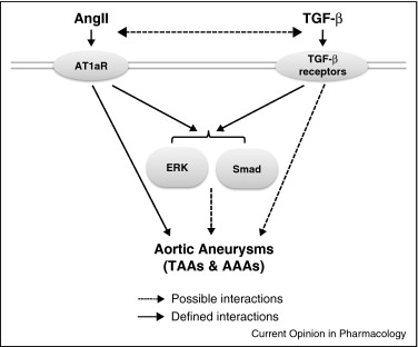
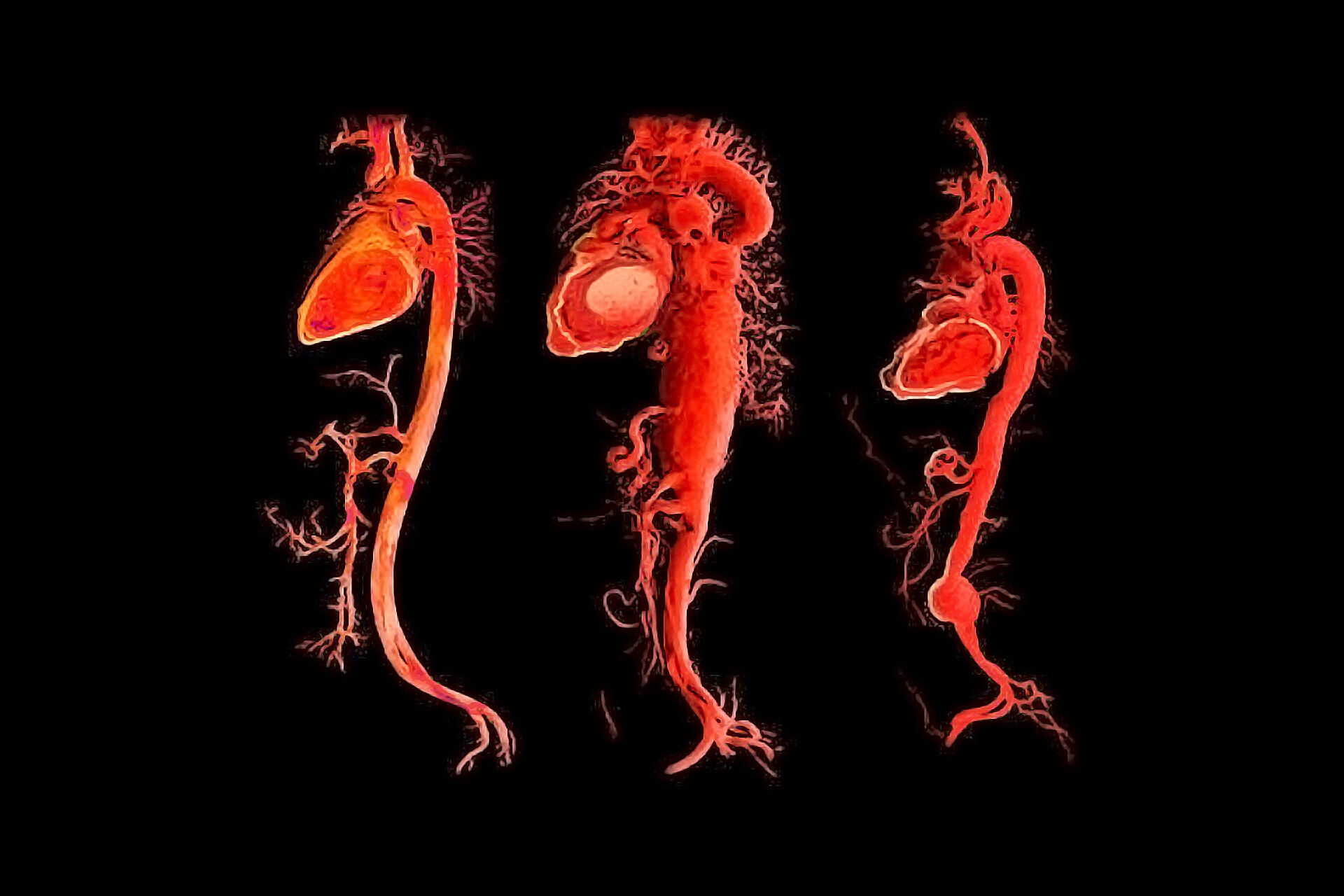
The involvement of Rac1 in various pathological aspects of muscular dystrophy positions it as a promising therapeutic target. By manipulating Rac1 signaling pathways, it may be possible to develop strategies that address not only inflammation and oxidative stress but also promote better muscle regeneration. Further research is needed to define the exact mechanisms of Rac1 in muscular dystrophies and its potential as a therapeutic target.
Research Assistant, exRNA Therapeutics
Are you curious about a rare condition affecting the skull and face? Watch this space for updates.
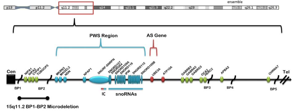
Auriculocondylar Syndrome (ACS) is an uncommon genetic condition that mostly impacts how the face develops, especially the jaw and ears. It is typically characterised by underdevelopment of the lower jaw (micrognathia) and a distinctive ear deformity known as the "question mark" ear. In some cases, people with ACS may also have a cleft palate, abnormal development of jaw muscles, and hearing loss. Genes related to face development, specifically GNAI3, PLCB4, or EDN1, are mutated in ACS. These genes are essential for the correct signalling during the development of the embryo's first and second pharyngeal arches, which give rise to the jaw and ear. The reported abnormalities are caused by gene mutations that interfere with proper signalling.
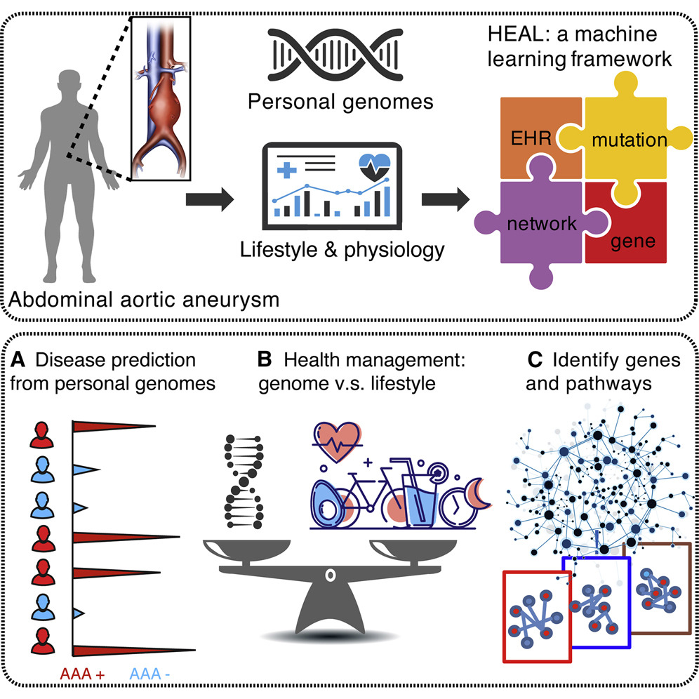
Guanine Nucleotide Binding Protein, Alpha Inhibiting Activity Polypeptide 3 is called GNAI3. This gene is specific for the inhibitory G protein's alpha subunit, which is essential for cellular signalling and inhibits the enzyme adenylyl cyclase. Numerous physiological functions, such as the control of cell proliferation and differentiation, depend on this inhibition.


heterotrimeric G-protein complex implicated in signal transduction, is encoded by the GNAI3 gene. Gαi3 is involved in a molecular switch mechanism that transfers signals from cell surface G-protein coupled receptors (GPCRs) to many intracellular signalling pathways. Its main function is to block adenylate cyclase, which lowers the amount of cyclic AMP (cAMP), a vital secondary messenger that controls hormone responses, cell division, metabolism, and other activities. The Gαi3 protein binds to and hydrolyzes GTP to modify signalling cascades, switching between active and inactive states. Gαi3 is involved in signal transduction, but it also affects other physiological functions such as endocytosis, vesicle trafficking, and cell proliferation.
The GNAI gene is crucial for several physiological functions. Appropriate regulation guarantees that cells react to outside stimuli precisely, preserving equilibrium in processes like:-

It is typified by unique deformities of the skull and face, like
Based on documented occurrences, ACS is thought to be an extremely uncommon hereditary condition with a frequency considerably below 1 in a million.
Research Assistant, exRNA Therapeutics
Alport Syndrome is a rare inherited disease that affects the kidneys, ears, and eyes. It is caused by mutations in genes responsible for producing type IV collagen, a crucial protein found in the basement membranes of the kidneys, cochlea (in the ear), and eyes. These mutations lead to progressive kidney failure, hearing loss, and, in some cases, eye abnormalities.

The kidneys are the most commonly affected organs in Alport Syndrome. The defective type IV collagen weakens the GBM, causing it to become progressively scarred and leaky. This results in early symptoms like hematuria (blood in urine) and eventually progresses to proteinuria (protein in urine). Over time, this damage can lead to a loss of kidney function, manifesting as CKD, and in severe cases, progresses to end-stage renal disease (ESRD), often requiring dialysis or kidney transplantation by early adulthood.
Diagnosis of Alport Syndrome is based on a combination of clinical findings, family history, and genetic testing to confirm mutations in the COL4A3, COL4A4, or COL4A5 genes. A kidney biopsy may also reveal characteristic abnormalities in the kidney’s basement membranes, supporting the diagnosis.
While there is currently no cure for Alport Syndrome, treatments are available to manage symptoms and slow the progression of kidney disease. Key strategies include:
Patients with Alport Syndrome benefit from comprehensive care, including support from nephrologists, audiologists, and ophthalmologists. Genetic counseling is important for families to understand inheritance patterns and risks for future generations.
Research into gene therapy and targeted molecular treatments for Alport Syndrome is ongoing. Early diagnosis, supportive care, and emerging treatments offer hope for better outcomes and improved quality of life for individuals with the disorder.
Alport Syndrome is a complex genetic condition with a profound impact on kidney function, hearing, and vision. Early diagnosis and proactive management are critical for delaying the progression of the disease and maintaining quality of life. Advances in genetic testing and treatment options continue to improve the outlook for those affected.
Research Assistant, exRNA Therapeutics
Alopecia universalis (AU) is an autoimmune condition marked by the total loss of hair on the scalp and body, representing a more severe form of alopecia areata, which typically causes localized patches of hair loss. The precise cause of AU remains unclear, but it is believed to involve the immune system mistakenly targeting hair follicles, leading to their destruction. This condition can significantly impact a person's emotional well-being and self-esteem, and while treatment options exist, outcomes can vary. The prevalence 1–5 cases per 10,000 people because it counts as a rare disorder .

Alopecia universalis (AU) is classified as an autoimmune disorder in which the immune system mistakenly attacks hair follicles. This condition is often associated with various genetic factors, particularly those involved in immune regulation. Key immune-related genes linked to AU include HLA-DRB1, the IL family, TYR, and interferon (IFN)-γ. These genetic components contribute to the immune response that triggers hair loss in affected individuals.
Alopecia universalis is an autoimmune condition characterized by the immune system attacking hair follicles, resulting in total body hair loss. While its primary causes are autoimmune in nature, various environmental factors may influence its onset and severity. Stress, whether physical or emotional, is often considered a potential trigger, although there is no definitive medical evidence linking it directly to alopecia universalis. Infections and exposure to certain chemicals or toxins might provoke autoimmune reactions in genetically predisposed individuals. Nutritional deficiencies and extreme environmental conditions can also impact hair and skin health.
Genetic factors play a crucial role in alopecia universalis, as it is a polygenic disease, meaning multiple genes contribute to its development. The condition can run in families, but both parents must pass on specific genes for it to be inherited, making its transmission complex and variable. Therefore, not every individual with alopecia universalis will have children affected by the condition.
Some other factor Hair loss caused by stress is termed telogen effluvium, which is temporary and not linked to immune or genetic issues. This type of hair loss typically occurs several months after significant stressors, such as illness or childbirth, and usually resolves on its own. Overall, while alopecia universalis involves a complex interplay of genetics and environmental factors, it remains distinct from stress-related hair loss.

Alopecia universalis currently lacks a definitive cure, but various treatment options can help manage symptoms or stimulate hair regrowth. In milder cases, the condition may stabilize without treatment. For more severe cases, options include:
Antisense oligonucleotide (ASO) therapy is being explored as a potential treatment for alopecia universalis (AU), a severe autoimmune-related hair loss condition. ASOs are synthetic strands designed to bind to specific mRNA, which may help modulate gene expression related to immune regulation and hair follicle development. By targeting pathways involved in immune dysregulation, ASOs could reduce the autoimmune attack on hair follicles and promote regrowth.
However, challenges remain, including effective delivery to hair follicles, ensuring specificity to prevent off-target effects, and addressing long-term safety and efficacy. While ASO therapy shows promise, further research and clinical trials are necessary to fully evaluate its potential in managing AU.
Alopecia universalis (AU) is a complex autoimmune condition characterized by total hair loss across the body. Its pathogenesis involves a dysregulated immune response targeting hair follicles, leading to significant psychological and emotional impacts for those affected. Current treatments, including topical therapies and immunosuppressants, often yield variable results. Emerging strategies, such as antisense oligonucleotide (ASO) therapies, offer hope by targeting the underlying molecular mechanisms of AU. However, further research is crucial to fully understand these therapies' effectiveness, safety, and long-term implications. Ultimately, a comprehensive approach that combines novel treatments with psychological support may improve outcomes for individuals living with AU.
Research Assistant, exRNA Therapeutics
Female infertility due to zona pellucida defect is a rare genetic disorder marked by the presence of abnormal oocytes that lack a functional zona pellucida. This protective glycoprotein layer is crucial for sperm binding and fertilization. In individuals with this condition, the absence or abnormality of the zona pellucida prevents normal fertilization processes, leading to infertility, Zona Pellucida defects is less than 1 in 1000000.

The Zona Pellucida Binding Protein (ZPBP) gene plays a critical role in sperm-egg interactions, and mutations in zona pellucida (ZP) encoding genes like ZP1, ZP2, and ZP3 can lead to zona pellucida defects and infertility, including conditions like empty follicle syndrome.
The zona pellucida-3 (ZP3) protein is crucial for the development of oocytes and gametes, ZP3 expression in oocytes is a key indicator of their fertilization capacity, Mutations in the ZP3 gene can disrupt normal zona pellucida formation, these mutations are associated with conditions like empty follicle syndrome.
Women with zona pellucida disorders often face challenges conceiving naturally or through assisted reproductive technologies like IVF.
Defective sperm-zona pellucida interactions are a major cause of low fertilization rates in IVF settings.

Genetic Testing- Analyzing genes related to the zona pellucida, such as ZP1, ZP2, and ZP3, to identify mutations.
Ultrasound Imaging- Using transvaginal ultrasound to examine ovarian follicles and the presence of any abnormalities.
Hormonal Testing- Measuring hormone levels to evaluate ovarian function and overall reproductive health.
Recent research underscores the importance of understanding the genetic basis of these defects to enhance fertility treatments.
Zona pellucida disorders are a significant focus within reproductive health, especially regarding female infertility. Understanding the genetic and functional roles of the zona pellucida can help develop targeted therapies and improve fertility treatment strategies.
Research Assistant, exRNA Therapeutics
Going ahead with knowing the gene involved, the next gene involved in causing Auriculocondylar Syndrome is PLCB4.
PLCB4 (phospholipase C beta 4) is the gene encoding an enzyme that is involved in cell signal transduction. PLCB4 is a member of the PLC family, which is in charge of transforming the signaling chemicals IP3 (inositol 1,4,5-trisphosphate) and DAG (diacylglycerol) from PIP2 (phosphatidylinositol 4,5-bisphosphate). PLCB4 is essential for several physiological processes. These chemicals facilitate the release of calcium from the endoplasmic reticulum activate protein kinase C, respectively, and set off following biological events.

PLCB4, mutations are the primary cause of Auriculocondylar Syndrome (ACS), a rare genetic disorder that affects the development of the face and lower jaw (mandible). One of the main characteristics of ACS is the abnormal development of the ears, mandible, and other craniofacial features. PLCB4's importance in this disease can be explained by its participation in the Edn1Dlx5/6 signaling pathway, which is crucial for normal craniofacial development. PLCB4 normally acts upon endothelin1 (Edn1), a signaling molecule crucial to controlling the development of the mandibular (lower jaw) arch. Edn1 activates PLCB4 via binding to its receptor, endothelin receptor type A, or EDNRA, which results in the production of DAG and IP3. Consequently, downstream transcription factors including Dlx5 and Dlx6 are activated, and calcium is released. The correct growth and patterning of craniofacial tissues depend on these variables. When the PLCB4 gene is mutated, the signaling function of the enzyme is disrupted. This leads to improper regulation of the calcium-dependent pathways responsible for the development of the jaw and ears. The development of ACS-specific features including undersized jaws (micrognathia) and abnormalities of the auricle (external ear) is caused by the disruption of the expression of crucial genes that control the size, shape, and structure of the jaw.
The PLCB4 gene encodes phospholipase C beta 4 (PLCβ4), a crucial element in intracellular signal transduction. This enzyme hydrolyzes the membrane phospholipid phosphatidylinositol 4,5bisphosphate (PIP2) to yield two important secondary messengers: inositol 1,4,5-trisphosphate (IP3) and diacylglycerol (DAG). In several signaling pathways ,those initiated by G-protein coupled receptors(GPCRs), PLCB4 is an essential component..When GPCRs activate PLCβ4, they produce IP3, which starts the release of calcium from intracellular stores, and DAG, which starts protein kinase C (PKC) activity. These molecules work together to regulate a variety of biological processes, including metabolism, growth, differentiation, and calcium-dependent signaling.
Auriculocondylar syndrome (ACS) is caused by mutations in the PLCB4 gene, which interfere with vital signaling pathways necessary for the normal development of face tissues, especially the lower jaws, and ears. The endothelin-1 (Edn1) signaling pathway depends on the enzyme phospholipase C beta 4 (PLCβ4) encoded by the PLCB4 gene. The development of craniofacial tissues, such as the mandible (lower jaw) and external ear, depends on this route. When PLCB4 is working correctly, it aids in the activation of downstream molecules that release calcium and activate important transcription factors like DLX5 and DLX6, which are essential for the development of the jaws and ears. Mutations in PLCB4, however, disrupt this process by resulting in defective communication that causes deformities such as auricular abnormalities and micrognathia, or tiny jaws.
Research Assistant, exRNA Therapeutics
Let's move to the last gene which is also involved in the cause of Auriculocondylar Syndrome, the name of the gene is EDN1.
Endothelin1 (ET1) is a potent vasoconstrictor peptide that plays a role in several physiological processes, most notably the cardiovascular system. It belongs to the endothelin family of peptides, which also includes endothelin 1 (ET-1),endothelin-2 (ET-2), and endothelin-3(ET-3).

Endothelial cells are the main source of the powerful vasoconstrictor peptide endothelin-1 (ET-1). Its primary activity is to cause smooth muscle cells in blood arteries to contract, which regulates blood pressure and vascular tone. In addition to its vasoconstriction function, ET1 is involved in inflammation, fibrosis, and cell proliferation. When overexpressed, these processes contribute to the pathophysiology of several disorders related to the heart and kidneys. Endothelin receptor type A mediates vasoconstriction and pro-inflammatory reactions, and endothelin receptor type B (ET_B), which can promote vasodilation through the production of nitric oxide and prostacyclin, are the two receptors that ET-1 binds to work. The intricate regulation of ET-1 includes feedback mechanisms, environmental cues, and transcriptional control. Shear stress, angiotensin II, hypoxia, and certain cytokines all promote the production. Its main objective is to induce contractions, which in turn control vascular tone and blood pressure. Conversely, it is downregulated by vasodilators such as atrial natriuretic peptide, nitric oxide, and prostacyclin. Furthermore, the production of ET-1 is regulated by the endothelin-converting enzyme (ECE), which converts Big ET-1 from an inactive precursor into an active version. Therefore, ET-1's activity and control are closely coordinated to maintain vascular homeostasis, with dysregulation contributing to diseases such as hypertension and atherosclerosis.
The powerful peptide endothelin1, which is produced by the EDN1 gene, is implicated in several physiological activities, chiefly those of the cardiovascular system. The primary action of endothelin-1 is vasoconstriction, which narrows blood vessels and lowers blood pressure. By interacting with the surface endothelin receptors (ET_A and ET_B) of vascular smooth muscle cells, ET-1 causes the cells to contract and expand blood arteries. Apart from its vasoconstrictive effects, ET-1 often causes inflammation, fibrosis, and cell division. These mechanisms help in tissue repair and wound healing. On the other hand, clinical problems may result from tissue remodeling and persistent vasoconstriction brought on by an excess of ET-1.
In patients with Auriculocondylar syndrome, mutations in the EDN1 gene alter the endothelin signaling pathway, leading to abnormal formation of these structures. The mutations especially impact ET-1's ability to regulate the development of the mandible, or lower jaw, and certain ear components. This leads to the characteristic features of the syndrome, which include malformed ears, small lower jaws, and other deformities of the face. The disruption is linked to altered interactions between endothelin receptors and their ligands, which are essential for controlling how facial tissues grow and differentiate in the developing embryo. Thus, abnormal signaling during craniofacial development resulting from mutations in EDN1 generates the distinctive features of Auriculocondylar syndrome.

According to most sources, ACS is classified as an uncommon ailment. This kind of hereditary problem typically affects less than 1 in 1,000,000 people. Given that ACS is caused by several genes (including EDNRA and GNAI3), the fraction of instances only resulting from EDN1 mutations would be a subset of this extremely uncommon occurrence.
Research Assistant, exRNA Therapeutics
Do you want to know about a disease in which lysosomes in excess amounts cause cellular dysfunction whose early symptoms are delayed speech and clumsy walking? If these symptoms are seen then this disease may be Aspartylglucosaminuria. So, let's read in detail about Aspartylglucosaminuria.
Aspartylglucosaminuria is a progressing disease comprising the neurodegenerative disease of which this condition is a type known as lysosomal storage disease. This disease is the result of incomplete aspartylglucosaminidase enzymes that are released from various body cells. This enzyme is important in the synthesis of glycoproteins. It breaks the bond between aspartate-glucosamine, which removes N-linked oligosaccharides. It helps to recycle and degrade the molecules in lysosomes. Consequently, aspartylglucosaminidase deficiency leads to undegraded Glycoasparagines in lysosomes in excess, which is a reason for cellular dysfunction. Aspartylglucosaminidase deficiency is due to the deletion of two base pairs in the aspartylglucosaminidase coding gene. Clinically, this ultimately leads to a deficiency of aspartylglucosaminidase which causes more bacterial infections than normal, and complications in the tissues with many blood vessels. Most patients suffering from AGU often die too early due to the effects of deadly diseases with the lungs being the major casualty. This condition is sporadic in the world and occurs mainly in Finland and 1 in million in the rest of the world.

Aspartylglucosaminuria (AGU) is a rare inherited disease found worldwide, but the majority of patients are from Finland due to a genetic founder effect. In Finland, 98% of cases are linked to the major AGUF variant, which involves two specific genetic changes. A minor variant is caused by the deletion of a two-base pair in the AGA gene (located on chromosome 4, specifically 4q34.3) which codes aspartylglucosaminidase enzyme and accounts for 1.5% of cases. There are more than 30 genetic variants of the AGA gene have been identified in people from various ethnic backgrounds, with about half being missense mutations. Other types of mutations include deletions, insertions, and complex changes. Specific mutations have been noted in certain populations, underscoring the genetic diversity in AGU. More details on the clinical implications of these variants will be covered later.
Aspartylglucosaminuria (AGU) affects the entire body which leads to intellectual disability, abnormal skeletal and connective tissue growth, and gait issues. Initial symptoms are delayed speech and clumsy walking, generally seen between 12-15 months. Children with AGU often experience frequent respiratory and ear infections and may require surgeries like tonsillectomies. Skeletal abnormalities include large head size, facial features like thick lips and a broad face, and short adult stature. Some patients may have scoliosis, a short neck, or knee problems. Brain imaging shows structural changes that indicate progressive neurological disease, and up to 28% of adults experience seizures. Early symptoms like diarrhoea and infections are common, while chronic arthritis affects a small percentage of patients. AGU patients may present with varying symptoms, such as hearing loss, muscular issues, or heart problems, depending on their background or other genetic conditions.

Areas with minimal knowledge regarding the disease Aspartylglucosaminuria have a high tendency of turning their patients into late diagnosis except in most cases some have been diagnosed with autistic individuals. Genetic testing for AGU is required and is ordered by clinical professionals and requires the use of blood or saliva samples. Testing involves sequencing of the AGA gene through NGS or Sanger sequencing techniques. Testing for AGU can be obtained in both commercial as well as academic laboratories. Accumulation of aspartylglucosamine in urine aspartylglucosaminuria (AGU) can be diagnosed by metabolic tests for AGU. Furthermore, blood sample analysis demonstrates that the activity of the enzyme aspartylglucosaminidase is lower than normal in aspartylglucosaminuria. Through biochemical tests or genetic testing, it is possible to determine the presence of AGU before birth, although it may need the use of cultured amniotic fluid cells. Such early clinical manifestations of AGU features might facilitate the diagnosis as delayed Speech, lack of coordination and a tendency to catch colds excessively. Diagnosis features are better developed as one grows including the conspicuous figure features and cognitive delays, some findings from brain magnetic scans also include poor differentiation of grey and white matter, poor myelination, and cyst formation.
Research Assistant, exRNA Therapeutics
MYH11 mutations are associated with a distinct vascular pathology that's potentially driven by insulin-like growth factor 1 (IGF-1) and angiotensin II (Ang II).
TAAD has been linked to several genes including : TGFBR1, TGFBR2, MYH11, FBN1, and ACTA2. MYH11 encodes the protein myosin-11, which is a component of the myosin heavy chain in smooth muscle. Mutations in the MYH11 gene affect the structure and assembly of the myosin thick filaments and have a dominant negative effect. Mutations in the MYH11 gene affect the structure and assembly of the myosin thick filaments and have a dominant negative

Thoracic aortic enlargement is an increasingly recognized condition that is often diagnosed on imaging studies performed for unrelated indications. The risk of unrecognised and untreated aortic enlargement and aneurysm includes aortic rupture and dissection which carry a high burden of morbidity and mortality. The etiologies underlying thoracic aortic enlargement are diverse and can range from degenerative or hypertension associated aortic enlargement to more rare genetic disorders. Therefore, the evaluation and management of these patients can be complex and requires knowledge of the pathophysiology associated with thoracic aortic dilation and aneurysm.

Anything that weakens your aorta’s walls can raise your risk for aneurysm formation. Besides atherosclerosis, known risk factors include: bicuspid aortic Valve disease, Family history of aortic aneurysm, Marfan syndrome.
The most common imaging technique for diagnosing a thoracic aortic aneurysm, this scan can show the size, location, and extent of the aneurysm.
Can provide information about the size of the aneurysm and the thoracic aorta. A transesophageal echocardiogram may be used if a standard echocardiogram doesn't provide enough information.
Uses magnetic fields and radio waves to create detailed images of the aorta and heart.
May show a convex contour of the right superior mediastinum, or an absence of the retrosternal airspace in the lateral chest x-ray.
Research Assistant, exRNA Therapeutics
Abdominal aortic aneurysm (AAA) is a multifactorial disease with a strong genetic component. Since first candidate gene studies were published 20 years ago, nearly 100 genetic association studies using single nucleotide polymorphisms (SNPs) in biologically relevant genes have been reported on AAA. The studies investigated SNPs in genes of the extracellular matrix, the cardiovascular system, the immune system, and signalling pathways. SNPs associated with AAA have already been identified in these large multicenter studies. One significant association was of a variant in a gene called CNTN3 which is located on chromosome 3p12.3.

An abdominal aortic aneurysm (AAA) is a swelling (aneurysm) of the aorta – the main blood vessel that leads away from the heart, down through the abdomen to the rest of the body.Large aneurysms are rare, but can be very serious. If a large aneurysm bursts, it causes huge internal bleeding and is usually fatal.The bulging occurs when the wall of the aorta weakens. Although what causes this weakness is unclear, smoking and high blood pressure are thought to increase the risk of an aneurysm.AAAs are most common in men aged over 65. A rupture accounts for more than 1 in 50 of all deaths in this group.
In most cases, an AAA causes no noticeable symptoms. However, if it becomes large, some people may develop a pain or a pulsating feeling in their abdomen (tummy) or persistent back pain. An AAA doesn’t usually pose a serious threat to health, but there’s a risk that a larger aneurysm could burst (rupture). A ruptured aneurysm causes massive internal bleeding, which is usually fatal. Around 8 out of 10 people with a rupture either die before they reach hospital or don’t survive surgery. The most common symptom of a ruptured aortic aneurysm is sudden and severe pain in the abdomen.
Among the 176 patients who had an unruptured aneurysm at the time of the initial ultrasound study, the cumulative incidence of rupture was 6 percent after 5 years and 8 percent after 10 years. However, the risk of rupture over five years was 0 percent for the 130 patients with an aneurysm less than 5 cm in diameter and 25 percent for the 46 patients with an aneurysm 5 cm or more in diameter. All 16 patients who had ruptures had aneurysms that were 5 cm or more in diameter at the time of the rupture.These population-based data challenge the clinical perception that aneurysms typically expand at a rate of 0.4 to 0.5 cm per year. Our data also suggest that for aneurysms less than 5 cm in diameter the risk of rupture is considerably lower than has been reported previously. However, the risk of rupture is substantial for aneurysms 5 cm or more in diameter.
Research Assistant, exRNA Therapeutics
Familial thoracic aortic aneurysm and dissection (TAAD) is a genetic condition characterised by the abnormal dilation or weakening of the thoracic aorta, which can lead to life-threatening dissections or ruptures. The genetics of familial thoracic aortic conditions involve several key aspects:
1. Hereditary Patterns:
A degenerative breakdown of collagen, elastin, and smooth muscle caused by ageing contributes to weakening of the wall of the artery.

Diagnosis: 1. Clinical Evaluation
Echocardiography
Research Assistant, exRNA Therapeutics
Chronic Kidney Disease (CKD) is a progressive condition that affects millions of people worldwide, leading to the gradual loss of kidney function over time. While CKD can arise due to various causes such as diabetes, hypertension, and autoimmune diseases, a specific subset of CKD cases is linked to genetic mutations. Among these are mutations in the COL4A3, COL4A4, and COL4A5 genes, which are central to the development of inherited kidney disorders like Alport Syndrome and Thin Basement Membrane Nephropathy (TBMN).

The COL4A3, COL4A4, and COL4A5 genes encode for different subunits of type IV collagen, a key structural protein found in the basement membranes of the kidneys, eyes, and inner ear. Basement membranes are thin, sheet-like structures that provide essential support for the tissues of these organs. In the kidneys, type IV collagen is especially critical for the structure and function of the glomerular basement membrane (GBM), which acts as a filtration barrier in the kidney's glomeruli.
The structure of type IV collagen consists of six different alpha chains (α1 to α6), with the α3(IV), α4(IV), and α5(IV) chains being particularly important in the kidney’s GBM. These chains form a network that maintains the integrity and function of the glomeruli, enabling the kidneys to filter blood and remove waste products efficiently.
1. COL4A5 Mutations and X-Linked Alport Syndrome
2. COL4A3 and COL4A4 Mutations and Autosomal Recessive Alport Syndrome
3. Thin Basement Membrane Nephropathy (TBMN)
Regular kidney function monitoring to assess disease progression.
The study of COL4A mutations has opened up new avenues for potential therapies, particularly gene therapy approaches that aim to replace the defective collagen chains. Research into medications that specifically target the molecular pathways involved in Alport Syndrome is ongoing, with the hope of preventing or delaying the onset of kidney failure in affected individuals. Additionally, animal models, particularly mice with COL4A3 or COL4A5 mutations, are helping researchers understand the progression of Alport Syndrome and test novel treatments. These models are crucial for developing future gene-editing or pharmacological interventions aimed at correcting the underlying genetic defects.
The COL4A3, COL4A4, and COL4A5 genes play a critical role in the structural integrity of the glomerular basement membrane. Mutations in these genes lead to various inherited kidney diseases, ranging from the mild hematuria of Thin Basement Membrane Nephropathy to the severe renal failure seen in Alport Syndrome. As genetic testing becomes more widely available, early diagnosis and management are improving outcomes for patients with these mutations. Ongoing research may one day lead to therapies that can correct the genetic defects and prevent the progression of CKD.
Research Assistant, exRNA Therapeutics
Chromosome 15q24 deletion syndrome at 24 position of chromosome 15 which is a rare condition and characterized by growth retardation, intellectual disability, distinctive facial features, as well as anomalies in the genitalia, skeleton, and digits. The condition is extremely rare, with an incidence of about 1 in 42,000 in the general population.The deletions associated with this syndrome range from 1.7 to 6.1 megabases (Mb) in size and includes CYP11A1, SEMA7A, CPLX3, ARID3B, STRA6, SIN3A and CSK genes which may cause various types of clinical features.

The common clinical features includes:
Diagnosis of 15q24 microdeletion syndrome primarily involves advanced genetic testing methods, as standard karyotyping often fails to detect such small deletions. The most common approach is genomic microarray analysis, which identifies copy number variations (CNVs) across the genome, specifically revealing deletions at the 15q24 locus. Array comparative genomic hybridization (aCGH) is also employed to confirm the presence and size of these deletions. Multiplex ligation-dependent probe amplification (MLPA) serves as a targeted screening tool for specific deletions and duplications. Alongside genetic testing, a thorough clinical evaluation is essential, where physicians look for characteristic features like growth retardation and intellectual disability. For at-risk pregnancies, prenatal testing can be conducted using amniotic fluid or chorionic villi samples to detect potential deletions before birth. Currently, there are no specific treatments that directly cure the genetic causes of 15q24 microdeletion syndrome. Treatment primarily focuses on managing symptoms and providing supportive therapies tailored to individual needs, such as occupational, physical, and speech therapies to tackle developmental delays and improve communication skills. While gene therapy has not yet been developed for this syndrome, recent studies are investigating the underlying genetic mechanisms. Researchers have identified key regions within the 15q24 locus and the genes involved that contribute to features of the syndrome. This knowledge may contribute to future gene therapy strategies aimed at restoring the function of affected genes or compensating for their loss, potentially leading to targeted treatments for specific symptoms or challenges. Ongoing research into gene dosage abnormalities and copy number variations (CNVs) may lead to innovative therapeutic options in the future.
Research Assistant, exRNA Therapeutics
Asherman’s syndrome is a rare, acquired, gynecological disorder of the uterus. It is characterized by the bonding of scar tissue that lines the walls of the uterus, which decreases the volume of the uterine cavity. The bonding of uterine scar tissue (intrauterine adhesions or synechiae) may occur as a result of surgical scraping or cleaning of tissue from the uterine wall (dilatation and curettage [D and C]), infections of the endometrium (e.g., genital tuberculosis), or other factors. Asherman’s syndrome can be severe (greater than 75% of the front and back walls of the uterus can fuse together) to moderate and mild, where only smaller portions of the uterine wall fuse together. This extra tissue creates less space inside the uterus. Women with Asherman’s syndrome may experience light or no periods and infertility. This condition can cause pelvic pain and abnormal uterine bleeding and can lead to fertility issues.
Uterine synechiae; Intrauterine adhesions; Infertility - Asherman
In most cases, it occurs in women who have had several dilatation and curettage (D&C) procedures.
A severe pelvic infection unrelated to surgery may also lead to Asherman syndrome.
Adhesions in the uterine cavity can also form after infection with tuberculosis or schistosomiasis. Uterine complications related to these infections are even less common.

The adhesions may cause:

Research Assistant, exRNA Therapeutics
An aortic aneurysm is a bulge that occurs in the wall of the body's main artery, called the aorta. The aorta carries blood from the heart to the body. Aortic aneurysms can occur anywhere in the aorta. They may be tube shaped or round.
Aortic aneurysms include:
An abdominal aortic aneurysm occurs along the part of the aorta that passes through the belly area.GSDMB gene was significantly upregulated in AAA macrophages, which was accompanied by robust non-canonical pyroptosis.
An thoracic aortic aneurysm occurs along the part of the aorta that passes through the chest cavity.Aortic aneurysm is considered a relatively rare but potentially life-threatening condition.

Abdominal aortic aneurysm (AAA): 1.4-3.9% in men, 0.5-1.7% in women (ages 50-79)
Thoracic aortic aneurysm (TAA): 0.5-1.1% in men, 0.2-0.6% in women (ages 50-79)
Abdominal Aortic Aneurysm: 2-4 per 100,000 person-years
Thoracic Aortic Aneurysm: 1-2 per 100,000 person-years
1. The majority of patients with an abdominal aortic aneurysm are asymptomatic and are diagnosed incidentally on imaging. When symptomatic, an abdominal aortic aneurysm usually presents with abdominal pain, flank pain, and thromboembolic events leading to limb ischemia. An untreated ruptured abdominal aortic aneurysm is fatal. The mortality rate of ruptured abdominal aortic aneurysms for patients reaching the hospital exceeds 50%.Hence, the identification of patients with an unruptured abdominal aortic aneurysm can be life-saving.

Many aneurysms develop without causing symptoms. Providers discover these aneurysms during a routine checkup or screening. If you’re at high risk of developing an aortic aneurysm — or have any aneurysm symptoms — your provider will do imaging tests. Imaging tests that can find and help diagnose an aortic aneurysm include:
If you have an unruptured aortic aneurysm, your provider will monitor your condition closely. If you have risk factors for developing an aortic aneurysm, your provider may also recommend regular screenings. Treatment aims to prevent the aneurysm from growing large enough to tear the artery or burst. For smaller, unruptured aneurysms, your provider may prescribe medications to improve blood flow, lower blood pressure or manage cholesterol. All can help slow aneurysm growth and reduce pressure on the artery wall.You can reduce your risk by maintaining a healthy lifestyle. This includes:
Research Assistant, exRNA Therapeutics
Amyotrophic lateral sclerosis (ALS), formerly known as Lou Gehrig's disease, is a neurological disorder that affects motor neurons, the nerve cells in the brain and spinal cord that control voluntary muscle movement and breathing.
As motor neurons degenerate and die, they stop sending messages to the muscles, which causes the muscles to weaken, start to twitch (fasciculations), and waste away (atrophy). Eventually, in people with ALS, the brain loses its ability to initiate and control voluntary movements such as walking, talking, chewing and other functions, as well as breathing.
Eventually, people with ALS will not be able to stand or walk, get in or out of bed on their own, use their hands and arms, or breathe on their own. Because they usually remain able to reason, remember, and understand, they are aware of their progressive loss of function.
The incidence of ALS is approximately 1-2.6 cases per 100,000 persons annually, whereas the prevalence is approximately 6 cases per 100,000.
As the disease progresses, muscle weakness and atrophy spread to other parts of your body. People with ALS may develop problems with:
Below we break down five of the most common misconceptions about ALS.
In reality, the majority of people diagnosed with ALS have no family history of the disease. About 90 percent of ALS cases are sporadic, meaning the cause or causes are unknown. The remaining 10 percent of ALS cases are familial and inherited through a mutated gene. In those families, there is a 50% chance that each offspring will inherit the gene mutation and may develop the disease.
Most people who develop ALS are between the ages of 40 and 70, with an average onset of 55. But there are many cases of people in their twenties and thirties who have been diagnosed with ALS. This “young onset” ALS typically refers to people younger than 45 years old and accounts for about 10% of all cases.
While not common, approximately 20% of individuals with ALS also develop frontotemporal degeneration (FTD) which is characterized by changes in personality, behavior and speech. Another rare form of ALS, known as ALS-parkinsonism-dementia complex (ALS-PDC) is characterized by signs and symptoms of ALS in addition to movement abnormalities (slow movements, stiffness and tremors) and loss of intellectual function. Cognitive and behavioral impairment in ALS can vary widely from individual to individual. Some people with ALS develop a symptom called the “pseudobulbar affect.” They may cry or laugh at inappropriate times or notice how once they start feeling an emotion, it is difficult to suppress it and not express it. Pseudobulbar affect is common in ALS and is the result of a brain reflex no longer working correctly. People with ALS can have pseudobulbar affect and no other cognitive, behavioral, or psychological symptoms.
People with ALS, even at an advanced stage, can still see, hear, smell, and feel touch. The nerves that carry feelings of hot, cold, pain, pressure, or even being tickled, are not affected by ALS. ALS is a disease that affects the parts of the nervous system that control voluntary muscle movement. Involuntary muscles, those that control the heart, GI tract, bowel and bladder function, and sexual functions are not directly affected by ALS.
From the initial symptoms to how the disease progresses, ALS can affect people very differently. Some people’s disease progresses very slowly, while others have changes that happen more quickly. Some individuals have severe involvement in one area before the disease spreads, while others experience rapid progression throughout their body. Some people may have severe weakness in one area, but little in others (e.g., unable to swallow but still able to walk and drive), and still others may demonstrate a similar severity of involvement in different areas. Eventually, ALS takes away the ability to walk, dress, write, speak, swallow, and breathe. How fast and in what order this occurs is very different from person to person.

Research Assistant, exRNA Therapeutics
Arterial tortuosity syndrome (ATS) is an extremely rare genetic disorder characterized by lengthening (elongation) and twisting or distortion (tortuosity) of arteries throughout the body.Affected arteries are prone to developing balloon-like bulges (aneurysms) on the wall of the artery, tearing (dissection), or narrowing (stenosis).
Arterial tortuosity syndrome is a connective tissue disorder. Connective tissues are the major components of the body forming skeleton, joints, skin, vessels and other organs. Connective tissues are characterized by the presence of cells included in an extracellular matrix network of a large variety of proteins (e.g., collagens), proteins bound to sugar chains of big dimension (proteoglycans) and sugars (hyaluronic acid, etc.
Arterial tortuosity syndrome exhibits autosomal recessive inheritance, and the responsible gene is located at chromosome 20q13.
The gene associated with arterial tortuosity syndrome is SLC2A10

ATS is a rare genetic disorder with an estimated prevalence of less than 1 in 1,000,000 live births.
It presents similarly to other collagen disorders with multiple multisystem abnormalities.
The condition predominantly affects the large and medium sized arteries.
The mechanism of this condition is apparently controlled by (or due to) the SLC2A10 gene.The molecular genetic pathogenesis finds that the SLC2A10 gene encodes the GLUT10 protein which is found in the nuclear membrane, or the endoplasmic reticulum, the latter of which GLUT10 transports DHA into.
The diagnosis of Arterial Tortuosity Syndrome is established in a person with generalized arterial tortuosity and positive molecular genetic testing that identifies a pathogenic mutation in the SLC2A10 gene. Generalized arterial tortuosity can be identified through the following:

The treatment of arterial tortuosity syndrome entails possible surgery for aortic aneurysm, as well as regular clinical surveillance including regular follow-up echocardiogram.
Research Assistant, exRNA Therapeutics
Bloom Syndrome (BSyn) is a rare genetic disorder that paints a complex picture of resilience and challenges. Named after Dr. David Bloom, who first described the condition in 1954, this syndrome is characterized by a series of clinical features that profoundly affect those who live with it. Understanding Bloom Syndrome requires not only a grasp of its medical implications but also an appreciation for the lives it touches.
Bloom Syndrome is caused by mutations in the BLM gene, which plays a crucial role in DNA repair. This genetic defect leads to increased susceptibility to various health issues, including:
The clinical manifestations of Bloom Syndrome are diverse:
Diagnosis typically involves genetic testing to identify pathogenic variants in the BLM gene. Management focuses on addressing symptoms and preventing complications:

Living with Bloom Syndrome involves navigating a landscape filled with unique challenges but also opportunities for growth and connection. Families affected by BSyn often develop close-knit communities that provide emotional support and share resources.
Bloom Syndrome serves as a poignant reminder of the complexities of genetic disorders. By fostering awareness and understanding, we can create a more inclusive environment for those affected by this condition. Their stories of resilience inspire us all to embrace our unique journeys, celebrating both the struggles and triumphs that define our lives. In sharing knowledge about Bloom Syndrome, we honor those who live with it, ensuring their experiences are recognized and valued within the broader tapestry of human diversity.
Research Assistant, exRNA Therapeutics
Bloom Syndrome (BSyn) is a rare genetic disorder that highlights the intricate relationship between our genes and health. Central to this condition is the BLM gene, which encodes a protein belonging to the RecQ helicase family. These proteins are crucial for maintaining the integrity of DNA, acting as the "caretakers of the genome." Understanding the mutations in the BLM gene provides insight into the challenges faced by individuals with Bloom Syndrome.
The BLM gene plays a vital role in several cellular processes:

Sister Chromatid Exchange: During cell division, sister chromatids—identical copies of chromosomes—can exchange small sections of DNA. This process, known as sister chromatid exchange (SCE), is essential for genetic diversity but can also be a response to DNA damage.

Mutations in the BLM gene lead to a deficiency or absence of functional BLM protein, resulting in several critical issues:
The consequences of these genetic mutations manifest in various ways:
Understanding the mutations in the BLM gene sheds light on the complexities of Bloom Syndrome. The interplay between genetic factors and health outcomes emphasizes the importance of ongoing research and awareness. By recognizing these connections, we can better support individuals living with this condition, ensuring they receive appropriate care and understanding. Bloom Syndrome serves as a poignant reminder of how our genetic makeup influences our lives, urging us to embrace both our vulnerabilities and strengths.
Research Assistant, exRNA Therapeutics

The structure of the BLM protein, which is encoded by the BLM gene associated with Bloom Syndrome. Here’s a breakdown of the key components depicted in the diagram:
Overall, this figure highlights the complex architecture of the BLM protein and its functional domains that are crucial for maintaining genomic stability. Understanding these structural components and mutations, like K695T, is essential for grasping how alterations in the BLM gene contribute to the manifestations of Bloom Syndrome, ultimately aiding in research and potential therapeutic approaches.
© eXRna Rare. All Rights Reserved 2024.
Designed by eXRna Rare
Distributed by eXRna Rare